ดาวน์โหลดงานนำเสนอ
งานนำเสนอกำลังจะดาวน์โหลด โปรดรอ

1
Interesting Case “ไข้ไอ ขาบวมจุดแดง”
พญ.ชนจันทร์ เพชรรัตน์ นพ.ปรีชา แสนยานุสิทธิ์กุล ยิ่งยง ชินธรรมมิตร์ 3/9/53

2
ผู้ป่วยชายไทยอายุ 37 ปี อาชีพ พนักงานไปรษณีย์ ภูมิลำเนา จ.ชัยนาท อาศัยที่ จ.กรุงเทพ 20 ปี สิทธิการรักษา เบิกได้ รับไว้ใน รพ.ศิริราช วันที่ 4/8/2553 อาการสำคัญ: ไข้ 10 วันก่อนมาโรงพยาบาล

3
อาการสำคัญ : ไข้ 10 วันก่อนมาโรงพยาบาล
ประวัติปัจจุบัน : 10 วันก่อนไข้สูงหนาวสั่นเป็นช่วงเย็นหลังอาบน้ำ กินพาราเซตตา มอลไข้ลด ไอแห้งๆ มีเจ็บหน้าอกด้านซ้ายเวลาไอ ไม่เหนื่อยหอบยัง ไปทำงานได้ ไม่มีปวดตามตัว-ปวดน่อง ไม่มีคลื่นไส้อาเจียน ไม่มี ตัว-ตาเหลืองไม่ปวดท้อง/ท้องเสีย ไม่มีเลือดออกผิดปกติที่ใด ไม่มีผื่น ไม่มีปัสสาวะแสบขัด 4 วันก่อน อาการไข้เท่า ๆ เดิม บางครั้งมีไอเสมหะปนเลือดติด กระดาษทิชชู ไปคลินิกได้ยา Ciprofloxacin (500) 1 tab bid Cetirizine (10) 1 tab hs, Paracetamol
 1 tab bid Cetirizine (10) 1 tab hs, Paracetamol.")
4
3 วันก่อน ไข้ ไอเท่าๆ เดิม เริ่มมีขาขวาบวมทั้งขา ปวดบริเวณน่องขวา มี แดงร้อน มีผื่นขึ้นขา 2 ข้าง เป็นจุดแดงเล็ก ๆ กระจาย ไม่คัน จึงมา โรงพยาบาล ประวัติเพิ่มเติม : ไม่มีประวัติเลือดออกง่ายผิดปกติมาก่อน เก็บหน่อไม้ในป่าที่จังหวัดชัยนาท 1 เดือนก่อน และช่วงเริ่มมีไข้แล้ว 1 วัน ไม่มีเบื่ออาหารหรือน้ำหนักลด ไม่มีประวัติมีผื่นแพ้แสง ผื่นที่หน้า ไม่มีผมร่วง ไม่มีปวดข้อมาก่อน ปัสสาวะออกดี ไม่มีฟอง ไม่มีปัสสาวะแดง

5
ประวัติอดีต ปฏิเสธโรคประจำตัว เดิมแข็งแรงดี ไม่เคยผ่าตัด
ประวัติครอบครัว ปฏิเสธโรคเลือด โรคมะเร็งในครอบครัว ประวัติส่วนตัว สูบบุหรี่ ดื่มเหล้าสัปดาห์ละครั้ง 5 ปี ปฏิเสธ unsafe sex IVDU การรับเลือดก่อนหน้านี้ ประวัติยา ไม่แพ้ยา กินยาที่ได้รับจากคลินิกได้ 3 วัน กินยาสมุนไพรต้มเองจากเปลือกไม้ 4 วันติดกันก่อนมา

6
Physical Examination Vital signs : T 38.2 C, PR 100 bpm, RR 20/min,
BP 103/65 mmHg, SpO2 94% RA GA : Good consciousness, no tachypnea, not pale, no jaundice, no sign of chronic liver disease, no clubbing of fingers SKIN : petechiae at both legs, no ecchymosis no malar rash, no eschar HEENT : no oral ulcer, no OC or OHL, no bleeding per gum Lymph nodes : no superficial lymphadenopathy

7
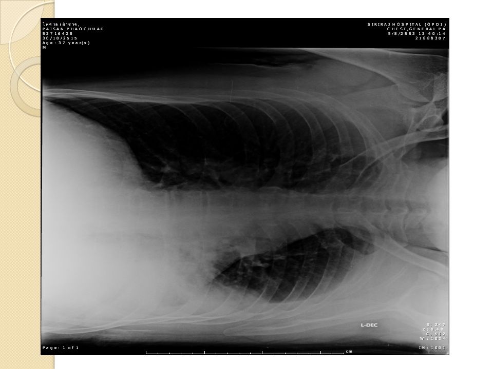
Physical Examination CVS: JVP 4 cm above sternal angle, no heaving, no thrill, normal S1S2, no murmur RS: trachea in midline, coarse crepitation and decreased breath sound LLL Abdomen: soft, not tender, no hepatosplenomegaly, shifting dullness negative NS: WNL

8
Physical Examination Extremities:
Left calf swelling with tenderness, warmth, mild ill-defined erythema Circumference leg Rt Lt cm Thigh Rt Lt cm Homan’s sign positive Lt leg Pulse FA , PA , DPA 2+ bilaterally capillary refill <2 seconds

9
Problem list Male 36 yr ไข้ ไอ จุดเลือดออก ขาซ้ายบวม Fever for 10 days
Cough, non-massive hemoptysis, left pleuritic chest pain for 10 days, and desaturation Petechiae both legs 3 days Left calf swelling and tenderness 3 days

10
Problem list Male 36 yr ไข้ ไอ จุดเลือดออก ขาซ้ายบวม Fever for 10 days
Cough, non-massive hemoptysis. pleuritic chest pain for 10 days, and desaturation Petechiae both legs 3 days Left calf swelling and tenderness 3 days Infectious Non infectious

11
Male 36 yr ไข้ ไอ จุดเลือดออก ขาซ้ายบวม
Infectious Systemic infection Rickettsial infection Leptospirosis Dengue infection Pulmonary infection Bacterial pneumonia Pulmonary tuberculosis Non infectious Pulmonary embolism with left leg DVT

13
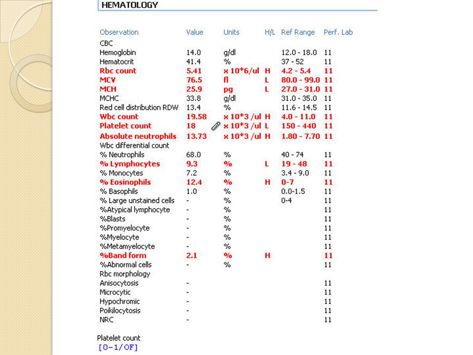
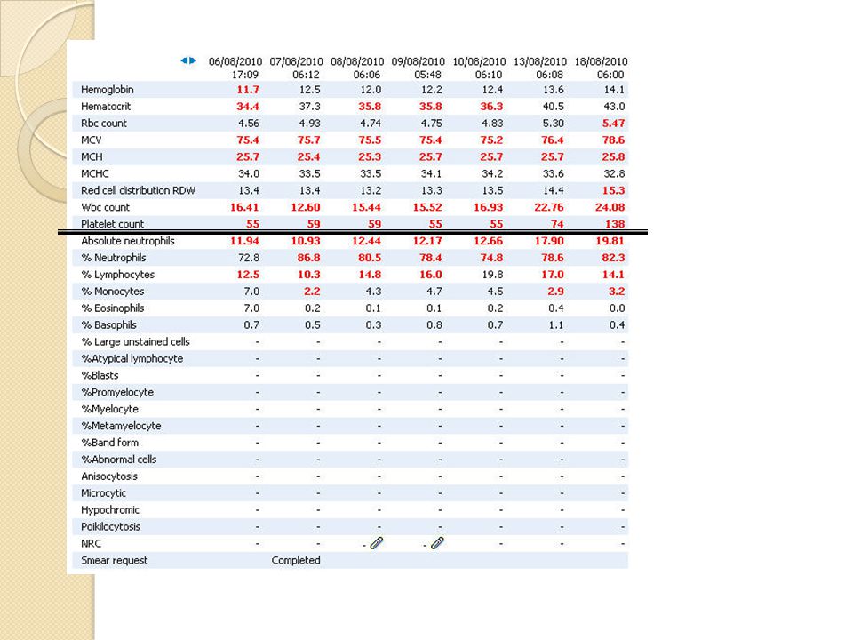
PBS

14
U/A

17
Problem list Male 36 yr ไข้ ไอ จุดเลือดออก ขาซ้ายบวม Fever for 10 days
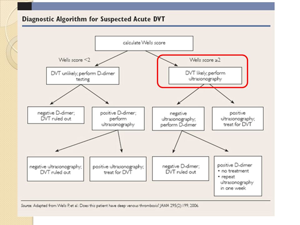
Cough, non massive hemoptysis for 10 days Petechiae both legs 3 days Left leg swelling 3 days DVT ? Cellulitis ?

18
Score 4 N Engl J Med 2003

20
Duplex scan Lt. Femoropopliteal vein thrombosis

21
Problem list Male 36 yr ไข้ ไอ จุดเลือดออก ขาซ้ายบวม
Fever for 10 days Cough ,non massive hemoptysis for 10 days Thrombocytopenia Left femoropopliteal vein thrombosis Intermittent fever for 10 days Cough ,non massive hemoptysis for 10 days Petechiae both legs 3 days Left leg swelling 3 days Infectious Non infectious : PE? Antiphospholipid syndrome Malignancy – solid/lymphoma with thrombocytopenia Infectionthrombocytopenia////DVT

22
Admit 5/8/53 Investigation Result Initial management Infectious
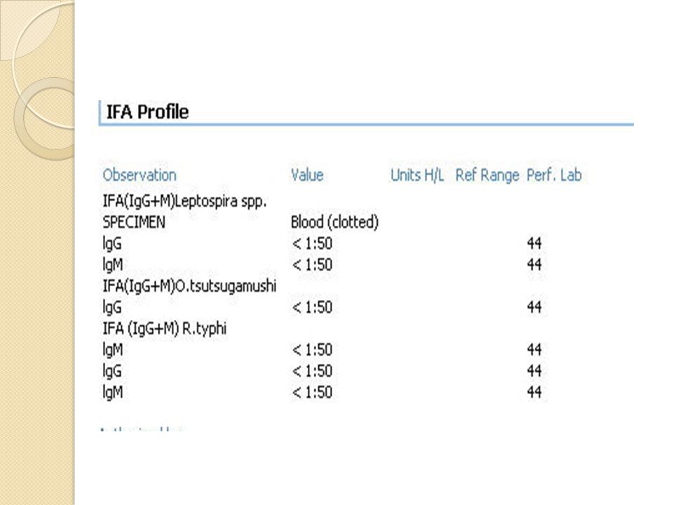
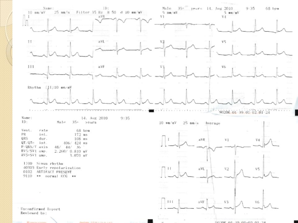
H/C x2 spps Sputum GS CS Sputum mycobacteria Dengue IgG IgM IFA O tsutsugamushi R typhi leptospirosis Pending เก็บไม่ได้ Ceftriaxone 2 g v OD Doxycycline(100) 1 tab o bid pc Non infectious PE? DVT Hypercoagulable? PT aPTT Lupus anticoagulant Anti B2 GP1 IgGIgM Anticardiolipin IgGIgM Heparin ? Platelet 18000? Thrombocytopenia Peripheral destruction /marrow disease Consult hemato STE inII ,III,aVF, cardiac enzyme no rising Consult cardiologist 22
 1 tab o bid pc. Non infectious. PE DVT. Hypercoagulable PT aPTT. Lupus anticoagulant. Anti B2 GP1 IgGIgM. Anticardiolipin IgGIgM. Heparin Platelet Thrombocytopenia. Peripheral destruction. /marrow disease. Consult hemato. STE inII ,III,aVF, cardiac enzyme no rising. Consult cardiologist. 22.")
23
Admit 5/8/53 Investigation Result Initial management Infectious
H/C x2 spps Sputum GS CS Sputum mycobacteria Dengue IgG IgM IFA O tsutsugamushi R typhi leptospirosis Pending เก็บไม่ได้ Ceftriaxone 2 g v OD Doxycycline(100) 1 tab o bid pc Non infectious PE? DVT Hypercoagulable? PT aPTT Lupus anticoagulant Anti B2 GP1 IgGIgM Anticardiolipin IgGIgM Heparin ? Platelet 18000? Thrombocytopenia Peripheral destruction /marrow disease Consult hemato STE inII ,III,aVF, cardiac enzyme no rising Consult cardiologist 23
 1 tab o bid pc. Non infectious. PE DVT. Hypercoagulable PT aPTT. Lupus anticoagulant. Anti B2 GP1 IgGIgM. Anticardiolipin IgGIgM. Heparin Platelet Thrombocytopenia. Peripheral destruction. /marrow disease. Consult hemato. STE inII ,III,aVF, cardiac enzyme no rising. Consult cardiologist. 23.")
24
8.5

25
High : Short term mortality in 30 days> 15%
European Heart Journal (2008) 29, 2276–2315
 29, 2276–2315.")
26
European Heart Journal (2008) 29, 2276–2315
 29, 2276–2315")
27
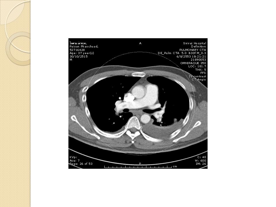
Investigation PE non high risk
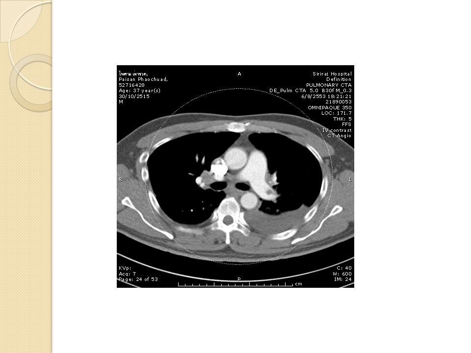
D-dimer-not recommended in high clinical probability normal result does not safely exclude High-probability ventilation–perfusion lung scintigraphy confirms PE (I, level A) SDCT or MDCT showing a segmental or more proximal thrombus confirms PE (I, level A) European Heart Journal (2008) 29, 2276–2315
 SDCT or MDCT showing a segmental or more proximal thrombus confirms PE (I, level A) European Heart Journal (2008) 29, 2276–2315.")
28
Ventilation–perfusion lung scintigraphy
OR CTA chest ?????

29
VQ scan vs CTA CTA Advantage over VQ scan
Speed Characterization of nonvascular structures, Detection of venous thrombosis. Caution in renal insufficiency, Ventilation–perfusion scan Diagnosis PE - high probability VQ scan in clinical probability intermediate to high False positive VQ scan* Prior pulmonary embolism Underlying cardiopulmonary disease 10% of smokers may have perfusion defect. N Engl J Med 2008 *Prog Cardiovasc Dis. 1994

32
ผลอ่าน

33
Problem list Male 36 yr ไข้ ไอ จุดเลือดออก ขาซ้ายบวม Fever for 10 days
Cough, non massive hemoptysis for 10 days Left femoropopliteal vein thrombosis Thrombocytopenia Pulmonary embolism with DVT

34
Management of PE Anticoagulation without delay - high or intermediate clinical probability of PE while workup (I, C) LMWH or fondaparinux - recommended initial Rx for most patients with non-high-risk PE (I, A) Unfractionated heparin (I, C) -High risk of bleeding - Severe renal dysfunction European Heart Journal (2008) 29, 2276–2315
 Unfractionated heparin (I, C) -High risk of bleeding. - Severe renal dysfunction. European Heart Journal (2008) 29, 2276–2315.")
35
? Patient Data --Thrombocytopenia????
Early Anticoagulation Is Associated With Reduced Mortality for Acute PE ? Patient Data --Thrombocytopenia???? Chest 2010;137;

36
PE …Thrombocytopenia Male 36 yr ไข้ ไอ จุดเลือดออก ขาซ้ายบวม ทีมแพทย์
Risk-benefit and Time anticoagulant Further inv and Rx thrombocytopenia ทีมแพทย์ ผู้ปวย ญาติ

37
DDx VTE and Thrombocytopenia
Anti-Phospholipid Syndrome - Primary - Secondary Malignancy : Solid tumor , Lymphoma - hypercoagulable state VTE - thrombocytopenia BM involvement, 2o ITP, CMT Paroxysmal nocturnal hemoglobinuria Heparin-induced thrombocytopenia/thrombosis (HITT) Two diagnoses eg, - VTE: Hereditary Hypercoagulable Disease eg, Protein C def , Protein S def, Antithrombin def - Thrombocytopenia: ITP, infection, drug, etc.
 Two diagnoses eg, - VTE: Hereditary Hypercoagulable Disease eg, Protein C def , Protein S def, Antithrombin def. - Thrombocytopenia: ITP, infection, drug, etc.")
38
Progress and managment
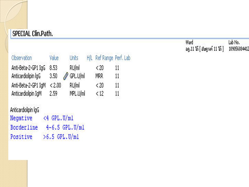
CTA pulmonary & CTV both legs Work up : hypercoagulable state -Lupus anticoagulant -anti-cardiolipin Ab -anti-β2GP I Ab -Protein C,Protein S -anti-thrombin III Ceftriaxone 2 gm iv O.D. Doxycycline(100)1x2 pc Paracetamal(500) 2 tab prn for fever q 4-6 hrs
1x2 pc. Paracetamal(500) 2 tab prn for fever q 4-6 hrs.")
39
Differential Diagnosis
Thrombocytopenia Peripheral destruction 1° or 2 °ITP ,DIC - Marrow Disease

40
Heparin iv in thrombocytopenia
Keep APTT ratio Keep Plt > 50,000 mm3 F/U Clinical bleeding/hemoptysis , CBC Protamine sulfate in hand

41
Day 1 2 3 4 5 6 pulse Temp Platelet 18,000 55,000 59,000 59,000 55,000
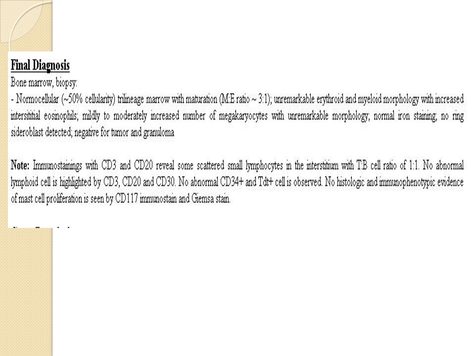
Pulse /min Temp C° Heparin iv pulse Ceftriaxone +doxycycline 120 40 BMA : reactive marrow ,no evidence of metastatic tumor ,lymphoma ,increased megakaryocytes Work up :anti-HCV,anti-HIV ,ANA -ve Temp 100 39 Warfarin 3 mg/day 80 38 -H/C –NG x 2sps -IFA for Scrub typhus,Leptospirosis - Dengue Ig G,IgM 60 37 50 36 40 35 BP mmHg Dexamethazone iv O2 Sat RA 90 % 90 % 90 % 95 % 97 % 99 % APTT Ratio 1.18 1.51 1.50 1.66 1.90 Hct 40 % 37 % 35.8 % 37.8 % 38.8 % Platelet , , , , ,000 41

43
Day 7 8 9 10 11 12 pulse Temp Platelet 55,000 74,000 BP mmHg CBC :
Pulse /min Temp C° CBC : Hb/Hct 13/40 Wbc 20,000 Plt 74,000 pulse Ceftriaxone +doxycycline 120 40 Heparin iv Temp 100 39 Warfarin 3 mg/day 80 38 60 37 50 36 Prednisolone 60 mg/day 40 35 BP mmHg O2 Sat RA 97 % 98% 98 % 98 % 98% 98% APTT Ratio 1.98 1.79 1.55 1.55 1.65 1.59 INR 1.79 2.8 Platelet , ,000 43

44
Day 13 14 pulse Temp Platelet 138,000 BP mmHg Warfarin 3 mg/day
Pulse /min Temp C° pulse 120 40 Temp 100 39 Warfarin 3 mg/day 80 38 60 37 50 36 Prednisolone 60 mg/day 40 35 BP mmHg O2 Sat RA 97 % 98% INR 2.36 Platelet ,000 44

50
Diagnosis Anti-phospholipid syndrome with ITP
Pulmonary embolism and DVT Immune thrombocytopenia Anti-phospholipid syndrome with ITP

51
Revised Classification Criteria for the Antiphospholipid Syndrome
At least one of the Clinical criteria At least one of the Laboratory criteria Miyakis S, J Thromb Haemost 2006; 4: 295–306.

52
Clinical Criteria Vascular thrombosis Pregnancy morbidity
arterial, venous, or small vessel thrombosis, in any tissue or organ For histopathologic confirmation, thrombosis should be present without significant evidence of inflammation in the vessel wall Pregnancy morbidity Miyakis S, J Thromb Haemost 2006; 4: 295–306.

53
Clinical Criteria Vascular thrombosis Pregnancy morbidity
>1 unexplained fetal death (GA >10 wk) >1 premature birth (GA <34 wk) due to (i) eclampsia or severe preeclampsia or (ii) placental insufficiency >3 unexplained consecutive spontaneous Abortions (GA <10 wk) Miyakis S, J Thromb Haemost 2006; 4: 295–306.
 >1 premature birth (GA <34 wk) due to (i) eclampsia or severe preeclampsia or (ii) placental insufficiency. >3 unexplained consecutive spontaneous Abortions (GA <10 wk) Miyakis S, J Thromb Haemost 2006; 4: 295–306.")
54
Laboratory criteria**
Anticardiolipin (aCL) antibody : IgG and/or IgM in medium or high titer (i.e. >40 GPL or MPL, or >99th percentile) Anti-2 glycoprotein-I antibody : IgG and/or IgM (>99th percentile) ** > 2 occasions at least 12 wk. apart Miyakis S, J Thromb Haemost 2006;4:295–306.
 antibody : IgG and/or IgM in medium or high titer (i.e. >40 GPL or MPL, or >99th percentile) Anti-2 glycoprotein-I antibody : IgG and/or IgM (>99th percentile) ** > 2 occasions at least 12 wk. apart. Miyakis S, J Thromb Haemost 2006;4:295–306.")
55
LA correlates better with thrombosis than aCL
Giannakopoulos B, et al. Blood 2009;113:985-94

56
Features associated with APS (non-criteria features of APS)
Heart valve disease Livedo reticularis Thrombocytopenia Nephropathy Neurological manifestation IgA aCL IgA anti-2 glycoprotein-I Antiphosphatidylserine Ab Antiphosphatidylethanolamine Ab Ab against prothrombin alone Ab against phosphatidylserine/prothrombin complex - MR/MS/AR/AS; Valve thickness >3 mm, Localized thickening involving the leaflet’s proximal or middle portion, Irregular nodules on the atrial face of the edge of the mitral valve, and/or the vascular face of the aortic valve. - In unselected APS patients, LR has been retrospectively correlated with aCL and arterial thrombosis, but not with anti-b2GPI or LA, venous thrombosis, or pregnancy morbidity - The committee consented that thrombocytopenia occurring in patients with persistent aPL, in the absence of clinical manifestations of APS, should be considered to be different from ITP: such patients have an increased thrombotic risk and require closer follow-up. - Thrombotic microangiopathy involving both arterioles and glomerular capillaries; Fibrous intimal hyperplasia involving organized thrombi with or without recanalization, Fibrous and/or fibrocellular occlusions of arteries and arterioles, Focal cortical atrophy, Tubular thyroidization (large zones of atrophic tubules containing eosinophilic casts) - In one small study of APS patients without SLE, long-term presence of LA is a risk factor for dementia (Evidence Level II). In SLE patients, persistent elevation of aPL is associated with cognitive dysfunction (Evidence Level I). Transverse myelopathy (TM) is a rare entity within APS. Limited data suggest that in the 1% of SLE patients who manifest TM, the latter is associated with aPL (Evidence Level IV) The IgA aCL are usually detected together with either IgG and/or IgM isotypes in patients with APS. In patients with collagen disease, IgA aCL associates with thrombocytopenia, skin ulcers and vasculitis, indicating a patient subgroup at risk for specific clinical manifestations (Evidence Level III), and it is highly prevalent in African–American SLE patients. IgA anti-b2GPI are the most frequently detected antibodies in patients in specific ethnic groups. A systematic review on antiprothrombin antibodies and risk of thrombosis in APS failed to reveal an association, irrespective of isotype, site and type of event, or presence of SLE. Both the sensitivity and specificity of aPS/PT are higher than those for aPT-A, whereas 95% of patients with aPS/PT are also LA positive, suggesting that aPS/PT can also serve as a confirmatory assay for LA (Evidence Level II); these results, however, only come from one study [128], and concerns regarding aPS/PT arise from multivalent antibody binding; the possibility of measuring antibodies against non-complexed phospholipids present in the sample needs to be excluded. Miyakis S, J Thromb Haemost 2006;4:295–306.
 - In one small study of APS patients without SLE, long-term presence of LA is a risk factor for dementia (Evidence Level II). In SLE patients, persistent elevation of aPL is associated with cognitive dysfunction (Evidence Level I). Transverse myelopathy (TM) is a rare entity within APS. Limited data suggest that in the 1% of SLE patients who manifest TM, the latter is associated with aPL (Evidence Level IV) The IgA aCL are usually detected together with either IgG and/or IgM isotypes in patients with APS. In patients with collagen disease, IgA aCL associates with thrombocytopenia, skin ulcers and vasculitis, indicating a patient subgroup at risk for specific clinical manifestations (Evidence Level III), and it is highly prevalent in African–American SLE patients. IgA anti-b2GPI are the most frequently detected antibodies in patients in specific ethnic groups. A systematic review on antiprothrombin antibodies and risk of thrombosis in APS failed to reveal an association, irrespective of isotype, site and type of event, or presence of SLE. Both the sensitivity and specificity of aPS/PT are higher than those for aPT-A, whereas 95% of patients with aPS/PT are also LA positive, suggesting that aPS/PT can also serve as a confirmatory assay for LA (Evidence Level II); these results, however, only come from one study [128], and concerns regarding aPS/PT arise from multivalent antibody binding; the possibility of measuring antibodies against non-complexed phospholipids present in the sample needs to be excluded. Miyakis S, J Thromb Haemost 2006;4:295–306.")
57
Thrombosis & Pregnancy morbidity
aPL 2GPI Activate endothelial cells & platelet ↓APC, ↓fibrinolysis Complement activation locally Thrombosis & Pregnancy morbidity

58
Detection of Lupus Anticoagulant Antibodies by in Vitro Coagulation Assays
The intrinsic coagulation pathway is initiated by contact activation on glass, silica, or kaolin (as in the activated partial-thromboplastin time [APTT], colloidal-silica clotting time [CSCT], and kaolin clotting time [KCT] assays), whereas the extrinsic coagulation pathway is initiated by the formation of a complex between tissue factor (TF) and factor VIIa (as in the dilute prothrombin time [dPT] assay). Russell’s viper venom directly activates factor X. Taipan, Textarin, and Ecarin snake-venom extracts directly activate prothrombin but have different cofactor requirements. Taipan venom activation of prothrombin requires phospholipid and calcium but not factor Va. Textarin activation of prothrombin requires phospholipid, calcium, and factor Va, whereas Ecarin activation of prothrombin is independent of cofactors and does not require phospholipid, calcium, or factor Va. Levine JS. NEJM 2002;346:
, whereas the extrinsic coagulation pathway is initiated by the formation of a complex between tissue factor (TF) and factor VIIa (as in the dilute prothrombin time [dPT] assay). Russell’s viper venom directly activates factor X. Taipan, Textarin, and Ecarin snake-venom extracts directly activate prothrombin but have different cofactor requirements. Taipan venom activation of prothrombin requires phospholipid and calcium but not factor Va. Textarin activation of prothrombin requires phospholipid, calcium, and factor Va, whereas Ecarin activation of prothrombin is independent of cofactors and does not require. phospholipid, calcium, or factor Va. Levine JS. NEJM 2002;346:")
59
Lupus Anticoagulant Test
Step 1 : Screening – Prolongation of coagulation in at least one phospholipid-dependent in vitro coagulation assay with the use of platelet-poor plasma Step 2 : Mixing study with normal pooled plasma – failure to correct the prolonged coagulation time Step 3 : Confirmation by shortening or correction of the prolonged coagulation time after the addition of excess phospholipid or platelets that have been frozen and then thawed Step 4 : Ruling out other coagulopathies with the use of specific factor assays if the confirmatory test is negative or if a specific factor inhibitor is suspected

60
LA1 / LA2 Ratio = Screening / Confirm Ratio ( a/c : b/d )
Fig. 2. Clotting times obtained with an LA positive test plasma or normal plasma (NP) with a low PL concentration (screening test) and a high PL concentration (confirmatory procedure). The PL concentration used in the ‘‘usual’’ APTT is indicated. The Lupus Ratio (a/b:c/d) is mathematically identical with the screening/confirm ratio (a/c:b/d).
 with a low PL concentration (screening test) and a high PL concentration (confirmatory procedure). The PL concentration used in the ‘‘usual’’ APTT is indicated. The Lupus Ratio (a/b:c/d) is mathematically identical with the screening/confirm ratio (a/c:b/d).")
61
Lupus Anticoagulant False positive False negative
Heparin contamination Specific anticoagulation factor antibody False negative Improper plasma preparation – platelet contamination = phospholipid Diluting effect of mixing studies – weak LA

62
A Meta-analysis Comparing LMWH – UFH in the Treatment of Venous Thromboembolism ARCH INTERN MED/VOL 160, JAN 24, 2000

63
0.47 (95% CI, ; P .06), but this did not reach statistical significance (Figure 4 Risk for heparin-induced thrombocytopenia with LMWH –UFH thromboprophylaxis: a meta-analysisBLOOD, 15 OCTOBER VOLUME 106,

64
Risk for heparin-induced thrombocytopenia with LMWH –UFH thromboprophylaxis: a meta-analysisBLOOD, 15 OCTOBER VOLUME 106,

งานนำเสนอที่คล้ายกัน