ดาวน์โหลดงานนำเสนอ
งานนำเสนอกำลังจะดาวน์โหลด โปรดรอ

1
วัตถุประสงค์ เมื่อสิ้นสุดการเรียนการสอนนักศึกษาสามารถอธิบาย
ส่วนประกอบของเนื้อเยื่อประสาทได้ถูกต้อง โครงสร้างของเซลล์ประสาทได้ถูกต้อง ชนิดและหน้าที่ของเซลล์ประสาทได้ถูกต้อง ชนิดและหน้าที่ของเซลล์ค้ำจุนประสาทได้ถูกต้อง คุณสมบัติของเนื้อเยื่อประสาทได้ถูกต้อง เส้นใยประสาท, โครงสร้างและชนิดของเส้นประสาทได้ถูกต้อง

2
จุดประสานประสาทได้ถูกต้อง
โครงสร้างและชนิดของปมประสาทได้ถูกต้อง โครงสร้างและชนิดของปลายประสาทได้ถูกต้อง โครงสร้างของระบบประสาทอัตโนมัติได้ถูกต้อง การเสื่อมและการงอกใหม่ของเส้นประสาท

3
พัฒนาการของระบบประสาท

5
การเจริญของ neuroectoderm
Ventricular zone (ependymal zone) Intermediate zone (mantle zone) Cortical plate (marginal zone) Geminal cell ใน ventricular zone เป็นเซลล์เริ่มต้น แบ่งตัวได้เซลล์ 3 ชนิดคือ - ependymoblast เจริญเป็น ependymal cell - gliobast เจริญเป็น supporting cell ยกเว้น microglia - postmitotic neurons (immature neurons)
 Intermediate zone (mantle zone) Cortical plate (marginal zone) Geminal cell ใน ventricular zone เป็นเซลล์เริ่มต้น แบ่งตัวได้เซลล์ 3 ชนิดคือ. - ependymoblast เจริญเป็น ependymal cell. - gliobast เจริญเป็น supporting cell ยกเว้น microglia. - postmitotic neurons (immature neurons)")
6
Neural crest cell เกิดระหว่างการ form neural tube เจริญเป็น
Dorsal root ganglion Autonomic ganglion Cranial nerve ganglion (V, VII, IX, X) Schwann cell and satellite cell Meninges Pigment cell Adrenal medulla Some skeletal and muscular component of the face
 Schwann cell and satellite cell. Meninges. Pigment cell. Adrenal medulla. Some skeletal and muscular component of the face.")
7
เนื้อเยื่อประสาท (nervous tissue)
เซลล์ประสาท (nerve cell or neuron) เซลล์ค้ำจุนประสาท (glia cell or neuroglia)
 เซลล์ค้ำจุนประสาท (glia cell or neuroglia)")
8
เซลล์ประสาท Cell body Cell processes

9
เซลล์ประสาท (neuron) 1. Cell body มีขนาดตั้งแต่ 5 -140 micron 1
เซลล์ประสาท (neuron) Cell body มีขนาดตั้งแต่ micron Nucleus มีขนาดใหญ่ รูปร่างกลม อยู่ตรงกลางเซลล์ มี nucleolus เด่น Cytoplasm มี organelles ดังนี้ Nissl (chromatophilic) body เป็น RER+ ribosome จำนวนมาก mitochondria มีมาก พบทั้งใน cell body และ cell processes Golgi body มีมากใน cell body แต่ไม่พบใน cell processes ไม่มี centriole microtubule and neurofilamentพบเป็นร่างแหใน cell body และทอดเป็นเส้นตามยาวใน cell processes มี inclusion ที่สำคัญคือ lipofuscin pigmentมีสีเหลือง เป็นผลจากการย่อย organelles โดย lysosome แต่ไม่ขับออกนอกเซลล์
 1.Cell body มีขนาดตั้งแต่ micron 1.1 Nucleus มีขนาดใหญ่ รูปร่างกลม อยู่ตรงกลางเซลล์ มี nucleolus เด่น 1.2 Cytoplasm มี organelles ดังนี้ Nissl (chromatophilic) body เป็น RER+ ribosome จำนวนมาก. mitochondria มีมาก พบทั้งใน cell body และ cell processes. Golgi body มีมากใน cell body แต่ไม่พบใน cell processes. ไม่มี centriole. microtubule and neurofilamentพบเป็นร่างแหใน cell body และทอดเป็นเส้นตามยาวใน cell processes. มี inclusion ที่สำคัญคือ lipofuscin pigmentมีสีเหลือง เป็นผลจากการย่อย organelles โดย lysosome แต่ไม่ขับออกนอกเซลล์")
10
Neuron structure

11
เนื่องจากเซลล์ประสาทมีรูปร่างได้ต่างๆ จึงเรียกชื่อเซลล์ประสาทตามรูปร่าง เช่น pyramidal cell, satellite cell, baskets cell, Purkinje’s cell เป็นต้น

12
2. Cell processes 2.1 Dendrite มีหลายแขนง มีขนาดใหญ่ สั้น เรียวเล็กลงอย่างรวดเร็ว แตกแขนงหลายครั้ง มี dendritic spine ที่ผิว 2.2 Axon มีเพียงแขนงเดียว เรียว ยาว ที่ตอนกลางพบ collateral branch ที่ปลายพบฝอยสั้นๆ (telodendria) ตรงปลายฝอยแต่ละอันพองออกเป็นกระเปาะเรียกว่า terminal bouton (axon terminal, synaptic knobs) axon hillock เป็นส่วนต้นต่อจาก cell body บริเวณนี้ไม่มี Nissl body
 ตรงปลายฝอยแต่ละอันพองออกเป็นกระเปาะเรียกว่า terminal bouton (axon terminal, synaptic knobs) axon hillock เป็นส่วนต้นต่อจาก cell body บริเวณนี้ไม่มี Nissl body.")
13
Dendrite

14
Axons Each neuron has only one axon but may possess a collateral branch All axons branches profusely at its terminal end to form more than 10,000 telodendria or terminal branches

15
Terminal bouton พบ vesicles เล็กๆ จำนวนมาก
ภายใน vesicles มี neurotransmiter เพื่อปล่อยออกมาเร้าเซลล์ประสาทตัวอื่นหรือเซลล์กล้ามเนื้อให้เกิดสัญญาณประสาท

16
การแบ่งชนิดของเซลล์ประสาท
1. แบ่งตามจำนวนแขนงที่ยื่นออกจากตัวเซลล์ 1.1 Pseudounipolar neuron 1.2 Bipolar neuron 1.3 Multipolar neuron

17
ชนิดของเซลล์ประสาทเมื่อแบ่งตามรูปร่าง

18
Pseudounipolar neuron
รุปร่างเป็นรูปตัว T แขนง peripheral process เป็น dendrite รับความรู้สึกเข้าไป แขนง central process เป็น axon ส่งความรู้สึกเข้าไปใน CNS พบใน spinal ganglion และ cranial ganglion (CN V, VII, IX and X)
")
20
Bipolar neuron มีแขนงยื่นออกจาก cell body สองแขนงทำหน้าที่เป็น dendrite หรือ axon พบในระบบประสาทรับความรู้สึกพิเศษ

21
Multipolar Neurons Multipolar neurons have more than two processes
Most common type in humans Major neuron of the CNS Most have many dendrites and one axon, some neurons lack an axon

22
2. ชนิดเซลล์ประสาทแบ่งตามหน้าที่
2.1Sensory or afferent neuron 2.2 Motor or efferent neuron 2.3 Association neuron or interneuron

23
Sensory Neurons Neurons that transmit impulses from sensory receptors in the skin or internal organs toward or into the CNS are called sensory or afferent neurons Virtually all primary sensory neurons of the body are unipolar

24
Motor Neurons Neurons that carry impulses away from the CNS to effector organs (muscles and glands) are called motor or efferent neurons Upper motor neurons are in the brain Lower motor neurons are in PNS

25
Motor neurons are multipolar and their cell bodies are located in the CNS (except autonomic)
Motor neurons form junctions with effector cells, signaling muscle to contract or glands to secrete

26
Interneuron or Association Neuron
These neurons lie between the motor and sensory neurons These neurons are found in pathways where integration occurs Confined to CNS Make up 99% of the neurons of the body and are the principle neuron of the CNS

27
Interneuron Neurons Almost all interneurons are multipolar
Interneurons show great diversity in the size and branching patterns of their processes

28
Interneurons The Pyramidal cell is the large neuron found in the primary motor cortex of the cerebrum The Purkinje cell from the cerebellum

29
Interneurons Stellate cells of the cerebellum
Granule cells of the cerebellum Basket cells of the cerebellum

30
ชนิดเซลล์ประสาทแบ่งตามความยาว axon
Golgi type I เป็นเซลล์ประสาทที่มี axon ยาวทำ หน้าที่เป็น tract ในสมองและไขสันหลัง Golgi type II เป็นเซลล์ประสาทที่มี axon สั้นๆทำ หน้าที่ประสานเชื่อมโยงภายในสมองและไขสันหลัง

31
เซลล์ค้ำจุนประสาท (neuroglia)
CNS Astrocytes or astroglia 2.Microglia Oligodendroglia Ependyma PNS Satellite cell Schwann cell

32
Astrocyte มีขนาดใหญ่ที่สุด รูปร่างคล้ายดาว มีแขนงมาก แบ่งออกเป็น2 ชนิด
มีขนาดใหญ่ที่สุด รูปร่างคล้ายดาว มีแขนงมาก แบ่งออกเป็น2 ชนิด Protoplasmic astrocyte Fibrous astrocyte เนื่องจาก intermediate filament มี glial fibrillary acidic protein (GFAP) มากดังนั้นจะเห็นจากการ ย้อมด้วย antibody ต่อ GFAP สังเกต: Perivascular feet และ perineural feet ทำหน้าที่รับและถ่ายสารอาหาร ควบคุมสิ่งแวดล้อมทางเคมีรอบๆ เซลล์ประสาท
 มากดังนั้นจะเห็นจากการ ย้อมด้วย antibody ต่อ GFAP. สังเกต: Perivascular feet และ perineural feet. ทำหน้าที่รับและถ่ายสารอาหาร ควบคุมสิ่งแวดล้อมทางเคมีรอบๆ เซลล์ประสาท.")
33
Oligodendroglia -มีขนาดเล็กกว่า astrocyte, มีแขนงน้อย
-สร้าง myelin sheath ให้กับ เส้นใยประสาทใน CNS

34
Microglia -มีขนาดเล็กที่สุด แขนงเล็ก ยาวหยิกงอ มีผิวเป็น ruffed border
-เกิดจาก bone marrow -ภาวะปกติพบน้อย แต่เมื่อเกิด nervous tissue injury จะเพิ่ม มากอย่างรวดเร็ว -มีหน้าที่ ทำลายสิ่ง แปลกปลอม เชื้อโรค เนื้อเยื่อ ที่ตาย (phagocytosis)
")
35
Ependymal cell -มีลักษณะคล้าย simple columnar epithelium
-บุผนัง ventricle ในสมองและ central canal ในไขสันหลัง -พบ cilia หรือ microvilli ที่ apical surface

36
Satellite cell: -พบในปมประสาท (ganglia) ล้อมรอบเซลล์ประสาทในปมประสาทนั้นๆ -ลักษณะเป็น cuboidal cell ขนาดเล็ก -กำเนิดจาก neural crest cell -ทำหน้าที่ควบคุมสิ่งแวดล้อมทางเคมีที่อยู่รอบๆ เซลล์ประสาทและป้องกันอันตรายต่อเซลล์ประสาท

37
Schwann cell ทำหน้าที่สร้าง myelin sheath โดยใช้เยื่อหุ้มเซลล์ของตัวเองพันล้อมรอบเส้นใยประสาท เรียกชั้นนอกที่มีแต่ cytoplasm และ nucleus ว่า neurilemma -นอกจากนี้ยังทำหน้าที่ช่วยซ่อมแซมเส้นประสาทเมื่อได้รับบาดเจ็บ

38
คุณสมบัติของเนื้อเยื่อประสาท
ความสามารถในการรับการกระตุ้น : สามารถสร้าง nerve impulse เมื่อมีการกระตุ้น ความสามารถในการเป็นสื่อนำ : เซลล์ประสาทสามารถนำกระแสประสาทส่งไปยังอวัยวะต่างๆ เพื่อการทำงานได้

39
1.Myelinated nerve fiber 2.Unmyelinated nerve fiber

40
Myelinated nerve fiber
เป็น white fibers มี myelin หุ้มโดยมี neurilemma เป็นชั้นนอก มี node of Ranvier อยู่ระหว่างปล้อง Myelin sheath เป็นสารไขมัน มีความจุไฟฟ้าต่ำ จึงเป็นฉนวน สัญญาณประสาทจะกระโดดข้ามส่วนนี้ไปเกิด action potential ที่ node of Ranvierการนำกระแสประสาทจึงไปได้เร็วกว่าที่ไม่มี myelin sheath หุ้ม เท่า

41
Neurilemma - เป็น Schwann ที่มีครบทั้ง cell membrane, cytoplasm และ nucleus

42
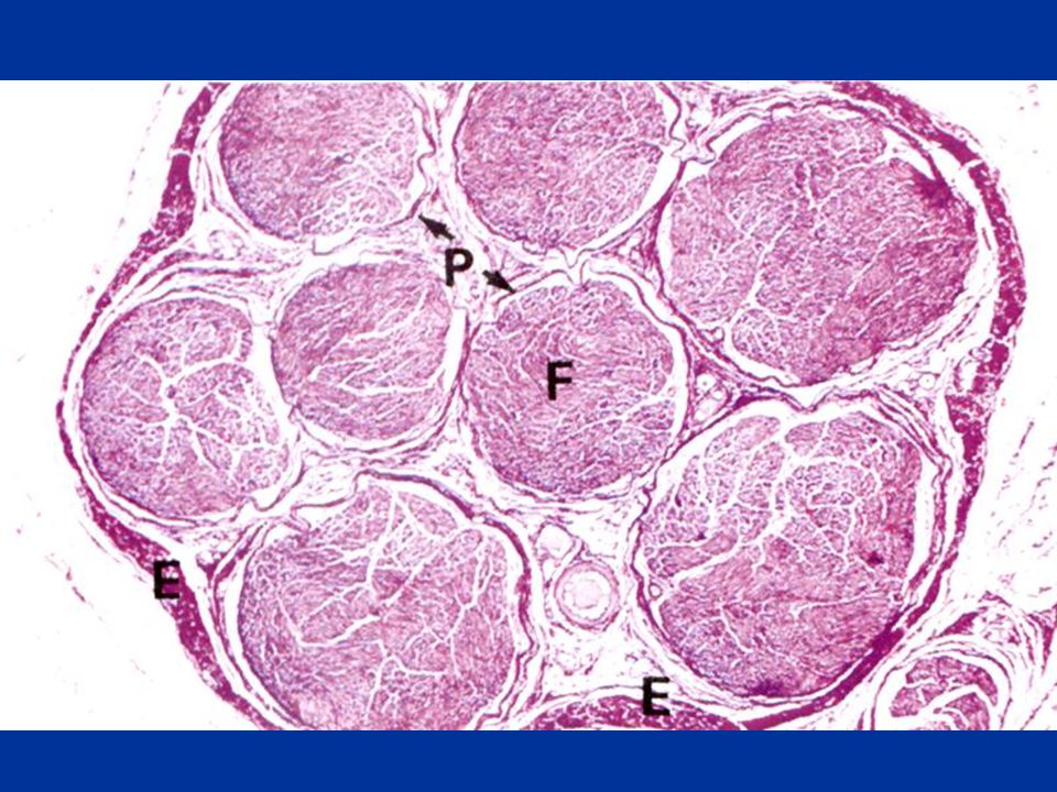
เส้นประสาท (nerve) ประกอบด้วยเส้นใยประสาทหลายเส้นรวมกัน
แต่ละเส้นใยถูกหุ้มด้วย endoneurium เส้นใยประสาทหลายๆ เส้นใยรวมเป็น bundle of nerve (fasciculus) หุ้มด้วย perineurium หลายๆ fasciculus รวมเป็น nerve หุ้มด้วยepineurium
 หุ้มด้วย perineurium. หลายๆ fasciculus รวมเป็น nerve หุ้มด้วยepineurium.")
46
ชนิดของเส้นประสาท แบ่งตามหน้าที่
เส้นประสาทนำคำสั่ง (motor nerve) นำคำสั่งจาก CNS ไปสู่อวัยวะต่างๆ เส้นประสาทรับความรู้สึก (sensory nerve) นำความรู้สึก จากตัวรับความรู้สึกไปยัง CNS เส้นประสาทผสม (mixed nerve) มีทั้งเส้นใยประสาทนำ คำสั่งและรับความรู้สึก
 นำคำสั่งจาก CNS ไปสู่อวัยวะต่างๆ. เส้นประสาทรับความรู้สึก (sensory nerve) นำความรู้สึก จากตัวรับความรู้สึกไปยัง CNS. เส้นประสาทผสม (mixed nerve) มีทั้งเส้นใยประสาทนำ คำสั่งและรับความรู้สึก.")
47
Table show classifying nerve fibers in peripheral nerve by diameter
Letter system Type of fiber Diameter micrometer Conduction Velocity, m/sec. General function A-alpha A-beta A-gamma A-delta B C 13-22 8-13 4-8 1-4 1-3 0.1-1 70-120 40-70 15-40 5-15 3-4 0.2-2 Alpha motor neuron, muscle spindle primary ending, Golgi tendon organs, touch Touch, kinesthesia, muscle spindle secondary ending Touch, pressure, gamma motor neurons pain, crude touch, pressure, temperature preganglionic autonomic pain, touch, pressure, temperature, postganglionic outom\nomic

48
Table show classifying nerve fibers in peripheral nerve by diameter
Roman system Type of fiber Diameter micrometer Conduction Velocity, m/sec. General function Ia Ib II III IV 12-20 11-19 5-12 1-5 0.1-2 70-120 66-114 20-50 4-20 0.2-3 muscle spindle primary ending Golgi tendon organs touch, kinesthesia, muscle spindle secondary ending pain, crude touch, pressure, temperature pain, touch, pressure, temperature

49
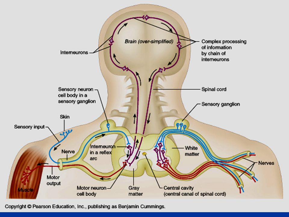
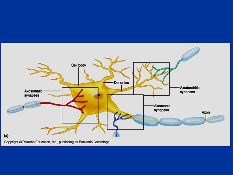
Synapse Synapse เป็นจุดประสานประสาท มีโครงสร้าง ประกอบด้วย
Presynaptic neuron Synaptic cleft Postsynaptic neuron

50
Synapses The site at which neurons communicate is called a synapse, a cell junction that mediates the transfer of information from one neuron to the next

52
ชนิดของ synapse Axodendritic synapse Axosomatic synapse
Axoaxonic synapse Dendrodendritic synapse

54
ปมประสาท (ganglion) เป็นกลุ่มเซลล์ประสาทที่อยู่นอกระบบประสาทส่วนกลาง มีเนื้อเยื่อเกี่ยวพันหุ้มล้อมรอบ มีโครงสร้างเป็นรูปไข่ ภายในมี cell body ของเซลล์ประสาท แต่ละเซลล์ล้อมรอบด้วย satellite cell

55
ชนิดของปมประสาท (Ganglion)
1.Sensory ganglion 1.1 Cranial ganglion Spinal ganglion 2.Autonomic ganglion Sympathetic ganglion Parasympathetic ganglion

56
Sensory ganglion มี cell body กลม ใหญ่ ภายในพบ fine Nissl’s body จำนวนมาก Nucleus อยู่ตรงกลางเซลล์ (concentric nucleus) แต่ละเซลล์ล้อมรอบด้วย satellite cells เป็น pseudounipolar neuron พบที่ dorsal root ของ spinal nerve ทุกเส้นและที่ ganglion ของ CNV, VII, IX และ X

57
Dorsal root ganglion

58
Autonomic ganglion

59
Autonomic ganglion Autonomic ganglion มีลักษณะกลมเป็นกระเปาะ
เซลล์ของปมประสาทอัตโนมัติเป็น multipolar neuron ภายในมี fine Nissl’s body เช่นเดียวกับเซลล์ของ Sensory ganglion Nucleus อยู่ขอบของเซลล์ (Eccentric nucleus) Sympathetic ganglion พบที่ paravertebral ganglion และ prevertebral ganglion Parasympathetic ganglion พบที่ terminal ganglion ซึ่งอยู่ที่ผนังอวัยวะที่ไปเลี้ยง
 Sympathetic ganglion พบที่ paravertebral ganglion และ prevertebral ganglion. Parasympathetic ganglion พบที่ terminal ganglion ซึ่งอยู่ที่ผนังอวัยวะที่ไปเลี้ยง.")
60
Sympathetic ganglion

61
Parasympathetic ganglion

62
Parasympathetic ganglion

63
ปลายประสาท (nerve fiber ending)
Motor nerve ending Sensory nerve ending

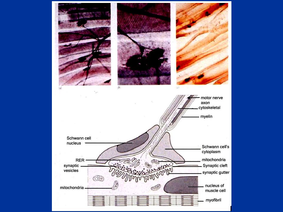
64
Motor nerve ending ปลายประสาท motor สิ้นสุดที่กล้ามเนื้อลาย เรียกว่า motor end plate หรือ neuromuscular junction (NMJ) Motor unit คือ กลุ่ม กล้ามเนื้อลายที่เลี้ยงด้วย motor neuron 1 ต้ว ก่อนสิ้นสุดจะเป็น unmyelinated nerve fiber

65
Motor nerve ending ในระดับ Em พบว่าปลาย axon พองใหญ่มี mitochondria และ vesicle ( Aº)จำนวนมาก ภายใน vesicle พบ acetylcholine ซึ่งเป็นสารสื่อ ประสาทที่สำคัญจำนวนมาก พบ synaptic celft กว้างประมาณ 500Aº ผนังของเซลล์กล้ามเนื้อพบเป็น รอยหยักโดยมีร่องเรียกว่า synaptic gutter

67
Motor nerve ending ที่กล้ามเนื้อหัวใจและกล้ามเนื้อเรียบ
Motor nerve ending ที่มาที่กล้ามเนื้อทั้งสองชนิดเป็นแบบไม่มี myelin sheath ปลายประสาทที่สิ้นสุดจะขยายใหญ่และอยู่ใกล้ชิดกับผนังกล้ามเนื้อนั้นๆ

68
ปลายประสาทรับความรู้สึก (sensory nerve ending
Unencapsulated nerve ending Encapsulated nerve ending

69
Unencapsulated nerve ending
Free nerve ending: รับความรู้สึกเจ็บปวด รับสัมผัส พบที่ epidermis, cornea, alimentary tract, gland, fascia, tendon, ligament, periosteum, joint capsule

70
Encapsulated nerve ending
1. Meissner’s corpuscle 2. Pacinian corpuscle 3. Ruffini’s corpuscle Krause’s end bulb 5. Muscle spindle 6. Golgi tendon organ

71
Pacinian corpuscle: รับความรู้สึกเกี่ยวกับแรงดัน ความสั่นสะเทือน การเคลื่อนไหวและท่าทางของข้อต่อ พบที่ subcutaneous ของฝ่ามือ ส้นเท้า หัวนม periosteum, mesentery, tendon,ligament

72
Meissner’s corpuscle: รับความรู้สึกสัมผัส แยกจุดสัมผัสสองจุด พบที่ dermal papilla โดยเฉพาะริมฝีปาก หัวนม นิ้ว อวัยวะเพศ

73
Ruffini’s corpuscle: รับรู้ต่อแรงดึง ความตึงตัวพบที่ dermis,joint capsule,CNT

74
Krause’s end bulb: were named after German anatomist Wilhelm Krause ( ).They have the ability to detect low-frequency vibration
.They have the ability to detect low-frequency vibration")
75
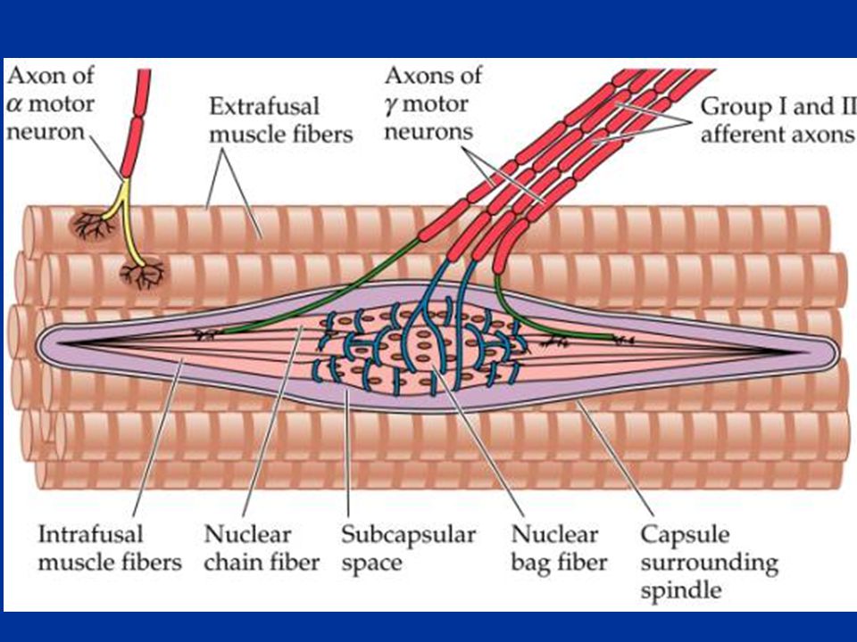
Muscle spindle เป็น modify ของเซลล์กล้ามเนื้อลายเปลี่ยนแปลงไปทำหน้าที่รับความรู้สึกเมื่อกล้ามเนื้อยึดตัวมาก เซลล์กล้ามเนื้อลายเล็กๆ ที่อยู่ภายในส่วนกลางของกล้ามเนื้อลายเปลี่ยนไปเป็นไม่มีลาย เรียกกลุ่มนี้ว่า intrafusal fibers ส่วนกล้ามเนื้อที่เหลือเรียกว่า extrafusal fibers เส้นประสาทสั่งการของ intrafusal fibers คือ gamma nerve fiberจาก gamma motor neuron ในไขสันหลัง เส้นประสาทสั่งการของ extrafusal fibers คือ alpha nerve fiberจาก alpha motor neuron ในไขสันหลัง

77
Intrafusal fibers Nuclear chain fibers เป็นเซลล์อยู่ตรงกลาง มีลักษณะเล็กยาว มีหลาย นิวเคลียส และเรียงกันแถวเดียว Nuclear bag fibers มีขนาดใหญ่กว่า นิวเคลียสเรียงตัวมากกว่า 1 แถว กลุ่มนี้อยู่รอบนอก nuclear chain fibers เส้นประสาท sensory ที่มารับความรู้สึกที่ nuclear bag และ nuclear chain ได้แก่เส้นประสาทที่เรียกว่า flower spray และ annulospiral fibers

78
Golgi tendon organ: พบที่รอยต่อระหว่างเอ็นกับกล้ามเนื้อลาย ถูกกระตุ้นเมื่อกล้ามเนื้อหดตัว ป้องกันกล้ามเนื้อหดตัวมากเกินไป

79
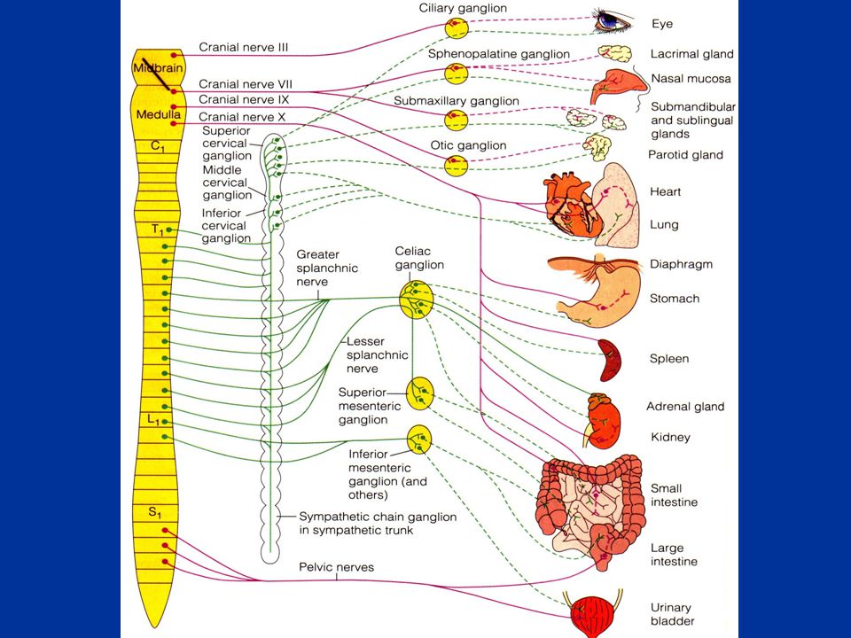
1. Sympathetic nervous system 2. Parasympathetic system
ระบบประสาทอัตโนมัติ (Autonomic nervous system, ANS) 1. Sympathetic nervous system 2. Parasympathetic system Preganglionic neuron Sympathetic: T1-L2 = Thoracolumbar Parasympathetic: Midbrain, Pons, Medulla and Sacral segment S2-S4 = Craniosacral Postganglionic neuron Sympathetic:Paravertebral ganglion (sympathetic chain) and prevertebral ganglion Parasympathetic: Terminal ganglion (intramural)
 1. Sympathetic nervous system 2. Parasympathetic system. Preganglionic neuron. Sympathetic: T1-L2 = Thoracolumbar. Parasympathetic: Midbrain, Pons, Medulla and. Sacral segment S2-S4 = Craniosacral. Postganglionic neuron. Sympathetic:Paravertebral ganglion (sympathetic chain) and. prevertebral ganglion. Parasympathetic: Terminal ganglion (intramural)")
81
ข้อสังเกต Adrenal medulla: ไม่มี postganglionic sympathetic fiber แต่จะหลั่ง epinephrine,norepinephrine ออกมาเอง postganglionic sympathetic fiber ที่ ผนังหลอดเลือดในกล้ามเนื้อลาย ต่อมเหงื่อ arrector pili m. ที่ผิวหนัง ให้ Ach ไม่มี parasympathetic fiber

83
sympathetic parasympathetic
ม่านตาหดตัว อัตราการเต้นของหัวใจลดลง หลอดลมหดตัว กระเพาะอาหาร ลำไส้บีบตัว หลั่งน้ำย่อยเพิ่มขึ้น กระเพาะปัสสาวะเพิ่มการบีบตัว อัณฑะแข็งตัว ม่านตาขยายตัว อัตราการเต้นของหัวใจเพิ่มขึ้น หลอดลมขยายตัว กระเพาะอาหาร ลำไส้บีบตัว หลั่งน้ำย่อยลดลง กระเพาะปัสสาวะลดการบีบตัว อัณฑะหลั่งน้ำกาม

84
1.Retrograde degeneration 2.Anterograte (Wallerian)degeneration
การเสื่อมของเส้นประสาท (Nerve degeneration) 1.Retrograde degeneration 2.Anterograte (Wallerian)degeneration
 1.Retrograde degeneration 2.Anterograte (Wallerian)degeneration.")
85
การงอกใหม่ของเส้นประสาท (Nerve regeneration)
")
งานนำเสนอที่คล้ายกัน


.>")
>")


 and Skeletal muscle contraction>")

>")