ดาวน์โหลดงานนำเสนอ
งานนำเสนอกำลังจะดาวน์โหลด โปรดรอ

1
Antepartum Fetal Health Assessment
Associate Professor Dr Atiwut Kamudhamas Department of Obstetrics and Gynecology Faculty of Medicine Thammasat University

2
Definition Fetal health assessment during the viable period before true labor pain

3
Assessment methods Fetal movement count Non stress test (NST)
Contraction stress test (CST) Fetal biophysical profile Doppler flow measurement Hormonal assay: Estriol, hPL
 Fetal biophysical profile. Doppler flow measurement. Hormonal assay: Estriol, hPL.")
4
Fetal movement count

5
Fetal movement count Principle Indication/ Contraindication Method
Interpretation Movement alarming signal Decrease fetal movement 24 hr then stop movement 8 hr before death All patients in 3rd trimester of pregnancy No contraindication Daily fetal movement record Cardiff count-to-10 See a doctor when movement< 10 times/ day

6
การนับการดิ้นของทารกในครรภ์
ข้อบ่งชี้ในการนับการดิ้นของทารก สตรีตั้งครรภ์ทุกรายที่การตั้งครรภ์เข้าสู่ไตรมาสที่ 3 หรือระยะที่ทารกเกิดมีชีพ(Viable period) การรับรู้ของสตรีตั้งครรภ์ต่อการดิ้นของทารก ส่วนต่างๆของทารกกระตุ้น subcutaneous tactile nerve ending ของผนังหน้าท้อง ทารกมีการเคลื่อนไหวในครรภ์ตั้งแต่ไตรมาสแรก การรับรู้การดิ้นของทารกครั้งแรก(quickening) : ~ GA wks ในครรภ์แรกจะรับรู้การดิ้นของทารกช้ากว่าครรภ์หลัง
 การรับรู้ของสตรีตั้งครรภ์ต่อการดิ้นของทารก. ส่วนต่างๆของทารกกระตุ้น subcutaneous tactile nerve ending ของผนังหน้าท้อง. ทารกมีการเคลื่อนไหวในครรภ์ตั้งแต่ไตรมาสแรก. การรับรู้การดิ้นของทารกครั้งแรก(quickening) : ~ GA wks. ในครรภ์แรกจะรับรู้การดิ้นของทารกช้ากว่าครรภ์หลัง.")
7
กลไกการดิ้นของทารก สัญญาณประสาทกระตุ้น Neuromuscular footplate ของกล้ามเนื้อโครงสร้างของทารก แหล่งกำเนิดสัญญาณประสาท Cerebral nerve root Spinal nerve root

8
Fetal behavioral states
State 1F : quiet sleep State 2F : active sleep (rapid eye movement) State 3F : quiet awake State 4F : active awake (FHR acceleration + vigorous body movement + REM)
 State 3F : quiet awake. State 4F : active awake (FHR acceleration + vigorous body movement + REM)")
9
รูปแบบการดิ้นของทารกที่อายุครรภ์ต่างๆ
อายุครรภ์ (สัปดาห์) รูปแบบการดิ้น 1- 8 8-12 13-16 16-20 20 ขึ้นไป ดิ้นรวดเร็วมาก เป็นจังหวะสั้นๆ ลักษณะกระตุก (Spastic) ดิ้นช้าลง ผสมผสานหลายรูปแบบมากขึ้น (Combined) ดิ้นมีแบบแผนพร้อมเพรียง (Coordinated type) ดิ้นคล้ายการขี่จักรยาน (Bicycling movement) ดิ้นแบบมีจุดประสงค์ (Purposeful like movement)
 รูปแบบการดิ้น ขึ้นไป. ดิ้นรวดเร็วมาก เป็นจังหวะสั้นๆ ลักษณะกระตุก (Spastic) ดิ้นช้าลง ผสมผสานหลายรูปแบบมากขึ้น (Combined) ดิ้นมีแบบแผนพร้อมเพรียง (Coordinated type) ดิ้นคล้ายการขี่จักรยาน (Bicycling movement) ดิ้นแบบมีจุดประสงค์ (Purposeful like movement)")
10
เกณฑ์การนับการดิ้นของทารกในครรภ์
การบันทึกการดิ้นของทารกใน 1 วัน (Daily fetal movement record) นับผลรวมจำนวนการดิ้นใน 12 ชั่วโมง แนะนำให้นับการดิ้นวันละ 3 ช่วง คือ 1 ชั่วโมงตอนเช้า ตอนเที่ยง และตอนเย็น ในแต่ละช่วงเวลา ถ้าดิ้นน้อยกว่า 3 ครั้งควรต่ออีก 1 ชั่วโมง ถ้าน้อยกว่า 10 ครั้ง ใน 12 ชั่วโมง ใน 2 วันติดกันถือเป็นอันตราย การนับทารกดิ้นจนครบ 10 ครั้ง (Cardiff count-to-ten) นับการดิ้นใน 12 ชั่วโมง ตั้งแต่หลังอาหารเช้า ( น.) ถ้าครบ 10 ครั้งให้หยุดนับได้ ถ้ายังไม่ครบให้นับต่อจนครบ 12 ชั่วโมง ถ้าครบ 12 ชั่วโมงทารกยังดิ้นไม่ถึง 10 ครั้ง ให้มาพบแพทย์
 นับผลรวมจำนวนการดิ้นใน 12 ชั่วโมง. แนะนำให้นับการดิ้นวันละ 3 ช่วง คือ 1 ชั่วโมงตอนเช้า ตอนเที่ยง และตอนเย็น. ในแต่ละช่วงเวลา ถ้าดิ้นน้อยกว่า 3 ครั้งควรต่ออีก 1 ชั่วโมง. ถ้าน้อยกว่า 10 ครั้ง ใน 12 ชั่วโมง ใน 2 วันติดกันถือเป็นอันตราย. การนับทารกดิ้นจนครบ 10 ครั้ง (Cardiff count-to-ten) นับการดิ้นใน 12 ชั่วโมง ตั้งแต่หลังอาหารเช้า ( น.) ถ้าครบ 10 ครั้งให้หยุดนับได้ ถ้ายังไม่ครบให้นับต่อจนครบ 12 ชั่วโมง. ถ้าครบ 12 ชั่วโมงทารกยังดิ้นไม่ถึง 10 ครั้ง ให้มาพบแพทย์")
11
Factor affecting fetal movement
GA Sleep awake cycle (20-40 min) Hypoxemia DFIU Uterine contraction Induction of labor Drug (alcohol, smoking, steroid) Chromosome abnormalities External stimuli Level of plasma glucose
 Hypoxemia. DFIU. Uterine contraction. Induction of labor. Drug (alcohol, smoking, steroid) Chromosome abnormalities. External stimuli. Level of plasma glucose.")
12
ความถี่การดิ้นต่างกันในแต่ละอายุครรภ์
Sadovsky,et al. 1979

13
ภาวะขาดออกซิเจน Acute hypoxemia Chronic hypoxemia
การดิ้นลดลงทันทีภายใน 10 นาที ใช้เวลา >30 นาที จึงจะมีการดิ้นที่เหมือนเดิม Chronic hypoxemia การดิ้นและการเต้นของหัวใจทารกจะเป็นปกติจากการปรับตัวทางสรีรวิทยา มี brain sparing effect

14
ปัจจัยที่เกี่ยวข้องกับการเคลื่อนไหวของทารกในครรภ์
ทารกเสียชีวิตในครรภ์ จะดิ้นน้อยลงมาก่อน ~ 24 hrs และมักหยุดดิ้น ~ 8 hrs ก่อนเสียชีวิต การเจ็บครรภ์ (labor) ทารกจะดิ้นมากขึ้นเมื่อมี uterine contraction การทำสูติศาสตร์หัตถการ มีการกระตุ้นบริเวณมดลูกและน้ำคร่ำ ทำให้ทารกในครรภ์ดิ้นมากขึ้น
 ทารกจะดิ้นมากขึ้นเมื่อมี uterine contraction. การทำสูติศาสตร์หัตถการ. มีการกระตุ้นบริเวณมดลูกและน้ำคร่ำ ทำให้ทารกในครรภ์ดิ้นมากขึ้น.")
15
Factor affecting perception of movement
Placental site Amniotic fluid volume GA Obesity Anxiety

16
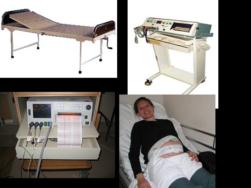
Management NST

17
Non stress test

18
Non stress test (NST) Principle Indication/ Contraindication Method
Interpretation FHR depend on the balance between sympathetic and parasympathetic activity Movement SympatheticFHR Hypoxia Parasym FHR Abnormal fetal movement count U/D (DM, HT, thyrotoxicosis) Postterm IUGR PROM Twins Preeclampsia Electronic fetal cardiotocography Semi-fowler (beware complication from supine hypotensive syndrome) 1. Reactive 2. Non reactive
 Postterm. IUGR. PROM. Twins. Preeclampsia. Electronic fetal cardiotocography. Semi-fowler (beware complication from supine hypotensive syndrome) 1. Reactive. 2. Non reactive.")
20
Reading NST Findings: 1. Baseline FHR ( bpm) 2. Variability 3. Abnormal pattern 4. Periodic change 4.1 Acceleration 4.2 Deceleration 5. Uterine contraction

21
Fetal heart rate acceleration
Increase FHR ≥ 15 beats per min and Persist > 15 sec <32 wks' : >10 bpm above baseline for >10 sec >32 wks' : >15 bpm above baseline for > 15 sec

22
การแปลผล NST Reactive Non-reactive
มี baseline FHR bpm และ Baseline variability 5-25 bpm และมี acceleration อย่างน้อย 2 ครั้งใน 20 นาที ถ้าไม่ครบตาม criteria ให้กระตุ้นทารกแล้วทำซ้ำอีก 20 นาที เพื่อเลี่ยง false non-reactive NST (mechanical/ vibroacoustic/biochemical) - No decelerations Non-reactive ไม่พบ acceleration หรือพบแต่ไม่ครบตามเกณฑ์วินิจฉัย reactive NST แสดงว่าทารกอาจอยู่ในภาวะไม่ปกติ แนะนำให้ทำการตรวจที่จำเพาะต่อไป เช่น CST, BPP
 - No decelerations. Non-reactive. ไม่พบ acceleration หรือพบแต่ไม่ครบตามเกณฑ์วินิจฉัย reactive NST. แสดงว่าทารกอาจอยู่ในภาวะไม่ปกติ แนะนำให้ทำการตรวจที่จำเพาะต่อไป เช่น CST, BPP.")
23
Reactive NST

24
Non-reactive NST

25
Reactive NST

26
Non-reactive NST with spontaneus deceleration

27
Non-reactive NST

28
Management Reactive F/U q 1 wk F/U 2-3 times/wk in DM type B-H, postterm, IUGR Nonreactive CST, BPP

29
Efficacy and effectiveness
High false positive Low positive predictive value High negative predictive value False negative NST 3.7% False positive NST 50% Negative predictive valve 92% Positive predictive valve 22%

30
Contraction stress test

31
Contraction stress test (CST)
Principle Indication/ Contraindication Method Interpretation Uterine contraction hypoxemia FHR Nonreactive NST (See next slide) 1.OCT 2.Nipple stimulation test 1.Negative 2.Positive 3.Suspicious 4.Hyperstimulation 5.Unsatisfactory
 1.OCT. 2.Nipple stimulation test. 1.Negative. 2.Positive. 3.Suspicious. 4.Hyperstimulation. 5.Unsatisfactory.")
32
Contraindications 1. Previous premature labour 2. Previous uterine surgery 3. Previous classical C/S 4. PROM 5. Placenta previa 6. Hydramnios 7. Incompetent cervix 8. Multiple gestation

33
Methods 1. oxytocin infusion 2. Nipple Stimulation
Start: 0.5 mU / min Titrate: increase 1 mU every 15 min 2. Nipple Stimulation Goal: contractions in 10 min Duration sec

34
Interpretation Negative Positive Suspicious Hyperstimulation
Unsatisfactory ไม่มี late deceleration และมี UC 3 ครั้งใน10 นาที พบ late deceleration มากกว่าครึ่งหนึ่งของจำนวน UC พบ late deceleration น้อยกว่าครึ่งหนึ่งของจำนวน UC มี UC ถี่กว่าทุก 2 นาที หรือ นานกว่า 90 วินาที หรือ 5 ครั้งใน10 นาทีและพบ late deceleration เส้นกราฟไม่มีคุณภาพเพียงพอ หรือ UC ไม่ดีพอ

35
Negative (reactive) CST
 CST")
36
Negative (reactive) CST
 CST")
37
Negative reactive CST

38
Negative nonreactive CST

39
Positive nonreactive CST

40
Hyperstimulation CST

41
Negative CST

42
Unsatisfactory CST

43
Unsatisfactory CST

44
การดูแลรักษาตามผล CST
Negative CST : ทารกอยู่ในสภาพปกติ แนะนำนับลูกดิ้นและตรวจซ้ำใน 1 สัปดาห์ Positive CST : ทารกอยู่ในสภาพพร่องออกซิเจน ช่วยเหลือโดย Intrauterine resuscitation และหยุด Oxytocin ทันที หลังจากนั้น นาทีให้ทำ CST ซ้ำ ถ้าผล Positive อีกครั้งควรสิ้นสุดการตั้งครรภ์ Suspicious CST : ทำการทดสอบ CST ซ้ำภายใน 24 ชั่วโมง Hyperstimulation : หยุด Oxytocin แล้วรอจนมี UC 3 ครั้งใน 10 นาทีจึงประเมินผลใหม่ Unsatisfactory CST : ตรวจซ้ำโดยจัดท่าสตรีและวาง transducer ในตำแหน่งที่เหมาะสม

45
Efficacy False negative CST 0.1% False positive CST 50%

46
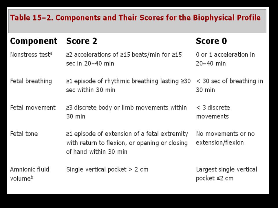
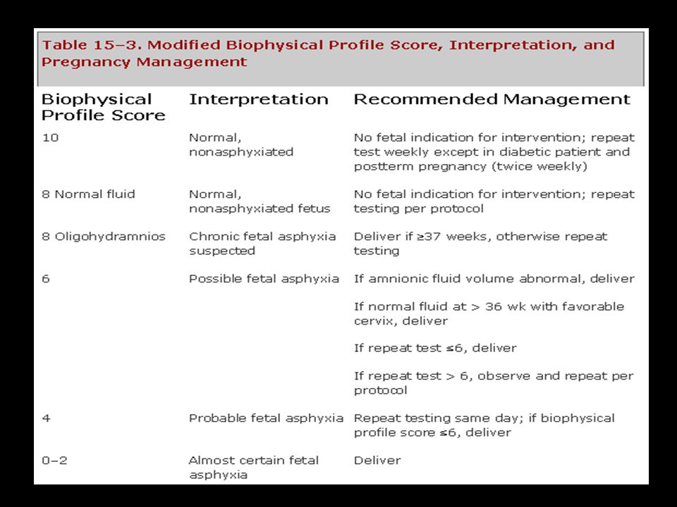
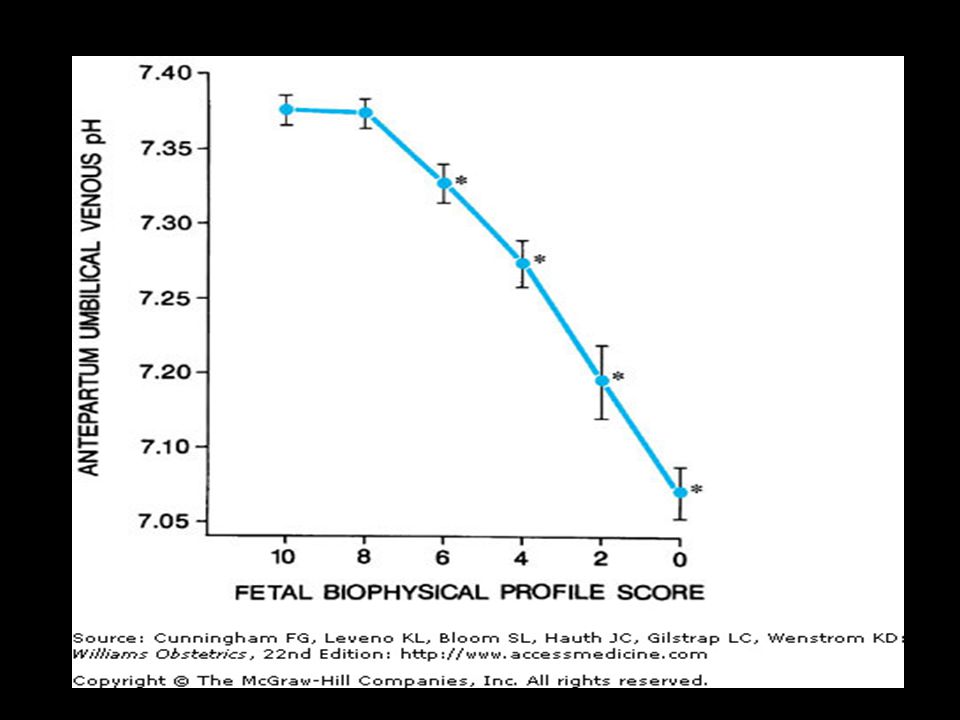
Fetal biophysical profile (BPP)
")
47
BPP Principle Indication/ Contraindication Method Interpretation
US + NST Nonreactive NST with contraindication of CST Back up surveillance NST Real time US American College of Obstetricians and Gynecologists (1999)
")
49
BPP scoring

51
Interpretation and management

53
Efficacy False negative BPP 0.007% False positive BPP 1%

54
Color Doppler measurement

55
Doppler flow measurement
การใช้คลื่นเสียงความถี่สูงแสดงภาพที่บอกความเร็วและปริมาณเลือดที่ผ่านหลอดเลือด ทำให้ทราบพยาธิสรีรวิทยาการไหลเวียนเลือดที่ทารกและรก ยังไม่มีข้อบ่งชี้ที่ชัดเจนในการทำ Doppler flow measurement เพราะผลยังมีความแปรปรวนค่อนข้างมาก นำมาใช้ติดตามสุขภาพทารกในครรภ์เสี่ยงสูงบางกรณี เช่น PIH IUGR Twin GDM

56
Vessels Umbilical artery Renal artery Uterine artery
Middle cerebral artery

57
Systolic/Diastolic ratio = A/B Resistance index = A-B/A
Pulsatility index = A-B/mean

58
Interpretation Umbilical systolic-diastolic ratio (S/D ratio)
Ratio >3 at GA > 37 weeks = abnormal More severe Absent end-diastolic flow Reversed end-diastolic flow UPI ↓ ความต้านทานของรกมากขึ้น การไหลเวียนเลือดช่วงDiastolic↓

59
Umbilical Artery Flow Systolic/Diastolic Ratio*
wks Mean Upper Limit

60
Normal

61
Absent end diastolic flow

62
Reverse diastolic flow

63
Hormonal assay

64
การสังเคราะห์เอสโตรเจนในสตรีตั้งครรภ์

65
การตรวจระดับ Estriol(E3) ในเลือดและปัสสาวะ
ระดับ E3 จะเพิ่มอย่างรวดเร็วในช่วงอายุครรภ์ wk และสูงคงที่ที่ 40 wk การตรวจทำได้ตั้งแต่อายุครรภ์ wk เป็นต้นไป การแปลผล E3 ที่อยู่ในช่วง 2SD ของแต่ละสัปดาห์ : ทารกปกติดี(Reassuring) E3 ที่ต่ำกว่าช่วง 2SD ของแต่ละสัปดาห์ : ไม่อาจรับประกันได้ว่าทารกปกติ (nonreassuring) พบผลบวกลวงสูงจากหลายปัจจัยที่รบกวนระดับ E3 สตรีตั้งครรภ์ใช้ยาสเตียรอยด์ ลดการดูดซึมที่ลำไส้สตรีตั้งครรภ์ เช่น กินยาปฏิชีวนะ
 E3 ที่ต่ำกว่าช่วง 2SD ของแต่ละสัปดาห์ : ไม่อาจรับประกันได้ว่าทารกปกติ (nonreassuring) พบผลบวกลวงสูงจากหลายปัจจัยที่รบกวนระดับ E3. สตรีตั้งครรภ์ใช้ยาสเตียรอยด์ ลดการดูดซึมที่ลำไส้สตรีตั้งครรภ์ เช่น กินยาปฏิชีวนะ.")
66
การตรวจระดับ Human placental lactogen(hPL)
ใช้ประเมินการทำงานของรกเป็นหลัก ข้อบ่งชี้ในการตรวจ : ไม่นิยมเนื่องจากผลบวกลวงและลบสูง Polypeptide hormone สร้างจาก syncytiotrophoblast เริ่มสร้างในสัปดาห์ที่ 3 หลังจากการตกไข่ ระดับสูงสุดเมื่อ GA wks ปริมาณสัมพันธ์กับขนาดของเนื้อรก ใช้ประเมินถึงการทำงานของรกเป็นหลัก ระดับปกติ คือ ภายในช่วง 2SD ของค่าเฉลี่ยในแต่ละGA

67
End of the session Thank you for your attention

งานนำเสนอที่คล้ายกัน


>")


>")

>")
>")
>")
>")
>")


