ดาวน์โหลดงานนำเสนอ
งานนำเสนอกำลังจะดาวน์โหลด โปรดรอ

1
การวัดความหนาแน่นของกระดูก Bone Densitometry
ดร.มนตรี ตั้งใจ ภาควิชารังสีเทคนิค คณะเทคนิคการแพทย์ มหาวิทยาลัยเชียงใหม่

2
เนื้อหานำเสนอ (Out line)
หลักการและเทคนิคในการวัดความหนาแน่นของกระดูก ปริมาณรังสีในการวัดความหนาแน่นของกระดูก โรคกระดูกพรุนและการตรวจ R2R

3
หลักการ I0 I I = I0 e -(M) โดย M คือ มวลสาร
คือค่าสัมประสิทธิ์การลดลงเชิงมวล(mass attenuation coefficient)
")
4
หลักการ I0 I I = I0 e -(M)
")
5
หลักการ I0 I I = I0 e -(M)
")
6
หลักการ I0 I I = I0 e -(M)
")
7
หลักการ 10 keV 40 keV ln (10/40) = - 0.9039 x M ln (I/I0) = - um x M
M = g/cm2 ln (I/I0) = - um x M ถ้า I0 = 40 keV I = 10 keV um = cm2 /g M = Mass per unit area
 = - um x M. ถ้า I0 = 40 keV. I = 10 keV. um = cm2 /g. M = Mass per unit area.")
8
หลักการ I0 (40 keV) = 40 I0 (70 keV) = 70 40 keV read out 70 keV
40 70 40 keV beam 70 keV beam X-ray tube I0 (40 keV) = 40 I0 (70 keV) = 70
 = 40. I0 (70 keV) = 70.")
9
หลักการ I040 = 40 Ist40 = 0.358 I070 = 70 Ist70 = 2.291 Soft Tissue
0.358 2.291 Soft Tissue (Fat+lean) Soft Tissue +bone I040 = 40 Ist40 = 0.358 I070 = 70 Ist70 = 2.291
 Soft Tissue +bone. I040 = 40 Ist40 = I070 = 70 Ist70 =")
10
หลักการ 0.08 1.96 I040 = 40 I070 = 70 Ist40 = Ist70 = 2.291 Ib40 = Ib70 = 1.96

11
หลักการ If mass attenuation coefficient ;
No tissue Soft issue Bone 40 keV read out I040 = 40 Ist40 = 0.358 Ib40 = 0.080 70 keV read out I070 = 70 Ist70 = 2.291 Ib70 = 1.960 If mass attenuation coefficient ; uf40 = uf70 = Rf = 1.211 uL40 = uL70 = RL= 1.421 ub40 = ub70 = Rb = 3.125 Need to calculate ust40 = ? ust70 = ? Rst = ?

12
หลักการ Rst = ln (I040 / Ist40) / ln (I070 / Ist70)
No tissue Soft issue Bone 40 keV read out I040 = 40 Ist40 = 0.358 Ib40 = 0.080 70 keV read out I070 = 70 Ist70 = 2.291 Ib70 = 1.960 Rst = ln (I040 / Ist40) / ln (I070 / Ist70) = ln (40 / 0.358) / ln ( 70 / 2.291) = ln (111.73) / ln (30.55) = / 3.419 = 1.379
 / ln (I070 / Ist70) = ln (40 / 0.358) / ln ( 70 / 2.291) = ln (111.73) / ln (30.55) = / =")
13
หลักการ No tissue Soft issue Bone 40 keV read out I040 = 40 Ist40 = 0.358 Ib40 = 0.080 70 keV read out I070 = 70 Ist70 = 2.291 Ib70 = 1.960 uf40 = uf70 = Rf = 1.211 uL40 = uL70 = RL= 1.421 ub40 = ub70 = Rb = 3.125 ust40 = ? ust70 = ? Rst = 1.379

14
หลักการ % Lean = [(Rst-RF)] / (RL-RF)] x 100
No tissue Soft issue Bone 40 keV read out I040 = 40 Ist40 = 0.358 Ib40 = 0.080 70 keV read out I070 = 70 Ist70 = 2.291 Ib70 = 1.960 % Lean = [(Rst-RF)] / (RL-RF)] x 100 = [( )] / ( )] x 100 = (0.168) / (0.21) x 100 = 0.8 x 100 = 80 %
![หลักการ % Lean = [(Rst-RF)] / (RL-RF)] x 100](http://slideplayer.in.th/slide/2177922/9/images/14/%E0%B8%AB%E0%B8%A5%E0%B8%B1%E0%B8%81%E0%B8%81%E0%B8%B2%E0%B8%A3+%25+Lean+%3D+%5B%28Rst-RF%29%5D+%2F+%28RL-RF%29%5D+x+100.jpg "No tissue. Soft issue. Bone. 40 keV read out. I040 = 40. Ist40 = Ib40 = keV read out. I070 = 70. Ist70 = Ib70 = % Lean = [(Rst-RF)] / (RL-RF)] x 100. = [( )] / ( )] x 100. = (0.168) / (0.21) x 100. = 0.8 x 100. = 80 %")
15
หลักการ % Lean = 80% %Fat = 100-%Lean %Fat = 100-80 %Fat = 20 %
No tissue Soft issue Bone 40 keV read out I040 = 40 Ist40 = 0.358 Ib40 = 0.080 70 keV read out I070 = 70 Ist70 = 2.291 Ib70 = 1.960 % Lean = 80% %Fat = 100-%Lean %Fat = %Fat = 20 %

16
Ust40 = (Lean Fraction)(UL40) + (Fat Fraction)(UF40)
No tissue Soft issue Bone 40 keV read out I040 = 40 Ist40 = 0.358 Ib40 = 0.080 70 keV read out I070 = 70 Ist70 = 2.291 Ib70 = 1.960 Ust40 = (Lean Fraction)(UL40) + (Fat Fraction)(UF40) = (0.80)(0.27) + (0.20)(0.23) = (0.216) + (0.046) = 0.262 Ust70 = (Lean Fraction)(UL70) + (Fat Fration)(UF70) = (0.80)(0.19) + (0.20)(0.19) = (0.152) + (0.038) = 0.19
(UL40) + (Fat Fraction)(UF40) = (0.80)(0.27) + (0.20)(0.23) = (0.216) + (0.046) = Ust70 = (Lean Fraction)(UL70) + (Fat Fration)(UF70) = (0.80)(0.19) + (0.20)(0.19) = (0.152) + (0.038) =")
17
หลักการ No tissue Soft issue Bone 40 keV read out I040 = 40 Ist40 = 0.358 Ib40 = 0.080 70 keV read out I070 = 70 Ist70 = 2.291 Ib70 = 1.960 uf40 = uf70 = Rf = 1.211 uL40 = uL70 = RL= 1.421 ub40 = ub70 = Rb = 3.125 ust40 = ust70 = Rst = 1.379

18
Mb = [Rst x ln (Ib70 / I070)] – ln (Ib40 / I040) Ub40 – (Ub70 x Rst)
No tissue Soft issue Bone 40 keV read out I040 = 40 Ist40 = 0.358 Ib40 = 0.080 70 keV read out I070 = 70 Ist70 = 2.291 Ib70 = 1.960 Mb = [Rst x ln (Ib70 / I070)] – ln (Ib40 / I040) Ub40 – (Ub70 x Rst) = [1.379 x ln(1.96 / 70)] – ln(0.80 / 40) 1.00 – (0.32 x 1.379) = 2.30 g/cm2
![Mb = [Rst x ln (Ib70 / I070)] – ln (Ib40 / I040) Ub40 – (Ub70 x Rst)](http://slideplayer.in.th/slide/2177922/9/images/18/Mb+%3D+%5BRst+x+ln+%28Ib70+%2F+I070%29%5D+%E2%80%93+ln+%28Ib40+%2F+I040%29+Ub40+%E2%80%93+%28Ub70+x+Rst%29.jpg "No tissue. Soft issue. Bone. 40 keV read out. I040 = 40. Ist40 = Ib40 = keV read out. I070 = 70. Ist70 = Ib70 = Mb = [Rst x ln (Ib70 / I070)] – ln (Ib40 / I040) Ub40 – (Ub70 x Rst) = [1.379 x ln(1.96 / 70)] – ln(0.80 / 40) 1.00 – (0.32 x 1.379) = 2.30 g/cm2.")
19
Mst = ln (Ib40 / I040)] – [Rb x ln (Ib70 / I070) (Rb x Ust70 ) – Ust40
No tissue Soft issue Bone 40 keV read out I040 = 40 Ist40 = 0.358 Ib40 = 0.080 70 keV read out I070 = 70 Ist70 = 2.291 Ib70 = 1.960 Mst = ln (Ib40 / I040)] – [Rb x ln (Ib70 / I070) (Rb x Ust70 ) – Ust40 = ln (0.08 / 40) – [3.125 x ln(1.96 / 70) (3.125 x 0.19 ) – 0.262 Mst (over the bone) = g/cm2
![Mst = ln (Ib40 / I040)] – [Rb x ln (Ib70 / I070) (Rb x Ust70 ) – Ust40](http://slideplayer.in.th/slide/2177922/9/images/19/Mst+%3D+ln+%28Ib40+%2F+I040%29%5D+%E2%80%93+%5BRb+x+ln+%28Ib70+%2F+I070%29+%28Rb+x+Ust70+%29+%E2%80%93+Ust40.jpg "No tissue. Soft issue. Bone. 40 keV read out. I040 = 40. Ist40 = Ib40 = keV read out. I070 = 70. Ist70 = Ib70 = Mst = ln (Ib40 / I040)] – [Rb x ln (Ib70 / I070) (Rb x Ust70 ) – Ust40. = ln (0.08 / 40) – [3.125 x ln(1.96 / 70) (3.125 x 0.19 ) – Mst (over the bone) = g/cm2.")
20
Mst = ln (Ist40 / I040)] – [Rb x ln (Ist70 / I070)
No tissue Soft issue Bone 40 keV read out I040 = 40 Ist40 = 0.358 Ib40 = 0.080 70 keV read out I070 = 70 Ist70 = 2.291 Ib70 = 1.960 Mst = ln (Ist40 / I040)] – [Rb x ln (Ist70 / I070) (Rb x Ust70 ) – Ust40 = ln (0.358 / 40) – [3.125 x ln(2.291 / 70) (3.125 x 0.19 ) – 0.262 Mst = 18.0 g/cm2
![Mst = ln (Ist40 / I040)] – [Rb x ln (Ist70 / I070)](http://slideplayer.in.th/slide/2177922/9/images/20/Mst+%3D+ln+%28Ist40+%2F+I040%29%5D+%E2%80%93+%5BRb+x+ln+%28Ist70+%2F+I070%29.jpg "No tissue. Soft issue. Bone. 40 keV read out. I040 = 40. Ist40 = Ib40 = keV read out. I070 = 70. Ist70 = Ib70 = Mst = ln (Ist40 / I040)] – [Rb x ln (Ist70 / I070) (Rb x Ust70 ) – Ust40. = ln (0.358 / 40) – [3.125 x ln(2.291 / 70) (3.125 x 0.19 ) – Mst = 18.0 g/cm2.")
21
% Lean of soft tissue (over bone) = 80%
No tissue Soft issue Bone 40 keV read out I040 = 40 Ist40 = 0.358 Ib40 = 0.080 70 keV read out I070 = 70 Ist70 = 2.291 Ib70 = 1.960 % Lean of soft tissue (over bone) = 80% % Fat of soft Tissue (over bone) = 20% So, ML (over bone) = x 0.80 = g/cm2 And MF (over bone) = x 0.20 = 2.99 g/cm2
 = 80% % Fat of soft Tissue (over bone) = 20% So, ML (over bone) = x 0.80 = g/cm2. And. MF (over bone) = x 0.20 = 2.99 g/cm2.")
22
% Lean of soft tissue (soft tissue) = 80%
No tissue Soft issue Bone 40 keV read out I040 = 40 Ist40 = 0.358 Ib40 = 0.080 70 keV read out I070 = 70 Ist70 = 2.291 Ib70 = 1.960 % Lean of soft tissue (soft tissue) = 80% % Fat of soft Tissue (soft tissue) = 20% So, ML (soft tissue) = 18 x 0.80 = 14.4 g/cm2 And MF (soft tissue) = 18 x 0.20 = 3.6 g/cm2
 = 80% % Fat of soft Tissue (soft tissue) = 20% So, ML (soft tissue) = 18 x 0.80 = 14.4 g/cm2. And. MF (soft tissue) = 18 x 0.20 = 3.6 g/cm2.")
23
Mb = 2.3 g / cm2 ML(over bone) = 11.96 g / cm2
No tissue Soft issue Bone 40 keV read out I040 = 40 Ist40 = 0.358 Ib40 = 0.080 70 keV read out I070 = 70 Ist70 = 2.291 Ib70 = 1.960 Mb = 2.3 g / cm2 ML(over bone) = g / cm2 MF(over bone) = 2.99 g / cm2 ML(soft tissue) = 14.4 g / cm2 MF (soft tissue) = 3.6 g / cm2 Total tissue weight= g
 = g / cm2. MF(over bone) = 2.99 g / cm2. ML(soft tissue) = 14.4 g / cm2. MF (soft tissue) = 3.6 g / cm2. Total tissue weight= g.")
24
เทคนิคในการวัดความหนาแน่นของกระดูก
การแบ่งประเภท 1. แบ่งตามการใช้งานของเครื่อง 1.1 เครื่องสำหรับวัดความหนาแน่นกระดูกบริเวณส่วนกลาง (Central devices) คือ บริเวณกระดูกสันหลัง (spine) กระดูกสะโพก (hip) 1.2 เครื่องสำหรับวัดความหนาแน่นกระดูกบริเวณส่วนระยางค์ (Peripheral devices) เช่น กระดูกข้อมือ (wrist) กระดูกส้นเท้า (heel)
 คือ บริเวณกระดูกสันหลัง (spine) กระดูกสะโพก (hip) 1.2 เครื่องสำหรับวัดความหนาแน่นกระดูกบริเวณส่วนระยางค์ (Peripheral devices) เช่น กระดูกข้อมือ (wrist) กระดูกส้นเท้า (heel)")
25
เทคนิคในการวัดความหนาแน่นของกระดูก
การแบ่งประเภท 2. แบ่งตามรูปแบบพื้นฐานของเครื่อง 2.1 แบบรังสีเอกซ์ (x-ray based) 2.2 แบบอัลทราซาวด์ (Ultrasound based)
 2.2 แบบอัลทราซาวด์ (Ultrasound based)")
26
เทคนิคในการวัดความหนาแน่นของกระดูก
การแบ่งประเภท 3. แบ่งตามโครงสร้างของกระดูก ( skeletal sites) 3.1 ส่วนกลาง (Central sites) 3.2 รยางค์ (Peripheral sites)
 3.1 ส่วนกลาง (Central sites) 3.2 รยางค์ (Peripheral sites)")
27
เทคนิคในการวัดความหนาแน่นของกระดูก
เครื่องวัดความหนาแน่นกระดูกบริเวณส่วนกลาง (Central densitometry devices) ได้แก่ 1. Dual-energy x-ray absorptiometry (DXA,DEXA) 2. Quantitative computed tomography (QCT)
 ได้แก่ 1. Dual-energy x-ray absorptiometry (DXA,DEXA) 2. Quantitative computed tomography (QCT)")
28
เทคนิคในการวัดความหนาแน่นของกระดูก
เครื่องวัดความหนาแน่นกระดูกบริเวณรยางค์ (Peripheral densitometry devices) ได้แก่ 1. Peripheral DXA (pDXA) 2. Single x-ray absorptiometry (SXA) 3. Peripheral QCT 4. Quantitive ultrasound (QUS) 5. Radiographic absortiometry (RA)
 ได้แก่ 1. Peripheral DXA (pDXA) 2. Single x-ray absorptiometry (SXA) 3. Peripheral QCT. 4. Quantitive ultrasound (QUS) 5. Radiographic absortiometry (RA)")
29
Gold standard Dual-energy x-ray absorptiometry (DXA, DEXA)
มีความแม่นยำสูง (excellent precision) ปริมาณรังสีน้อย (effective dose : 1-3 Sv) นำไปใช้ในการศึกษาทางระบาดวิทยา ติดผลการรักษาและการตอบสนองของยา เป็นที่รู้จักกันดี (well known) ใช้งานง่าย
 ปริมาณรังสีน้อย (effective dose : 1-3 Sv) นำไปใช้ในการศึกษาทางระบาดวิทยา. ติดผลการรักษาและการตอบสนองของยา. เป็นที่รู้จักกันดี (well known) ใช้งานง่าย.")
30
DXA scanner

31
What does the x-ray beam look like?

32
Pencil Beam Vs fan beam Pencil Beam Fan Beam Line acquisition
Multiple detector Faster acquisition Point acquisition Single detector Slower acquisition

33
Precision Error and Trueness Error
Central DXA Precision Error Trueness Error Spine % % Femur % 6% Forearm 1% 5% Total Body 1% 3% Genant HK et al. J Bone Miner Res. 1996; 11(6):707
:707.")
34
Precision Error and Trueness Error
Peripheral DXA Precision Error Trueness Error Forearm 1-2 % % Calcaneus 1-2% % Hand 1-2% 5% Genant HK et al. J Bone Miner Res. 1996; 11(6):707
:707.")
35
Effective Radiation Dose ( Sv) for DXA
Scan Mode QDR1000/4000 QDR4500/delphi DPX-L Prodigy Pencil Beam Fan Beam Pencil Beam Fan Beam Spine (L1-L4) Femur Forearm Total body Blake GM et al, The evaluatuon of osteoporosis,1999 Mammography 450 Chest x-ray Lateral lumbar x-ray 700 L-spine + T-spine Bonnick S and Lewis LA: Bone densitometry for technologistes Humana Press 2006 Bone densitometry course technologist course syllabus and associated reading materials 2008 (ISCD)
 Femur Forearm Total body Blake GM et al, The evaluatuon of osteoporosis,1999. Mammography 450 Chest x-ray Lateral lumbar x-ray 700 L-spine + T-spine Bonnick S and Lewis LA: Bone densitometry for technologistes Humana Press Bone densitometry course technologist course syllabus and associated reading materials 2008 (ISCD)")
36
What is Bone Densitometry?
This test takes bone scans to check density or thickness of bone. Bone Density Testing Is Done For Three Reasons To diagnose osteoporosis To predict fracture risk To monitor therapy

37
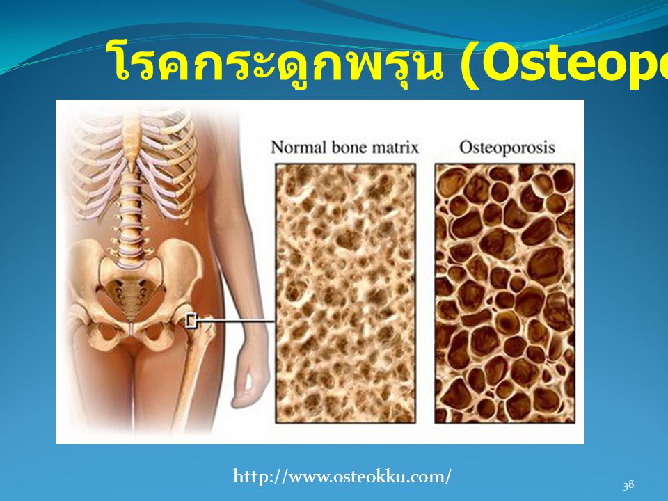
โรคกระดูกพรุน (Osteoporosis)
Osteoporosis is a disease of bones that leads to an increased risk of fracture. In osteoporosis the bone mineral density (BMD) is reduced, bone microarchitecture is deteriorating
 is reduced, bone microarchitecture is deteriorating.")
38
โรคกระดูกพรุน (Osteoporosis)
39
Risk Factors Age As people age, their risks for osteoporosis increase. Aging causes bones to thin and weaken All women over age 65 and All men over age 70 Postmenopausal women under age 65 with one or more risk factors for osteoporosis Any man or woman over age 50 who has suffered a fracture

40
Risk Factors Body Type Osteoporosis is more common in people who have a small, thin body frame and bone structure. Family History People whose parents had a history of fractures may be more likely to have fractures Dietary Factors Calcium and vitamin D deficiencies are important factors in the risk for osteoporosis

41
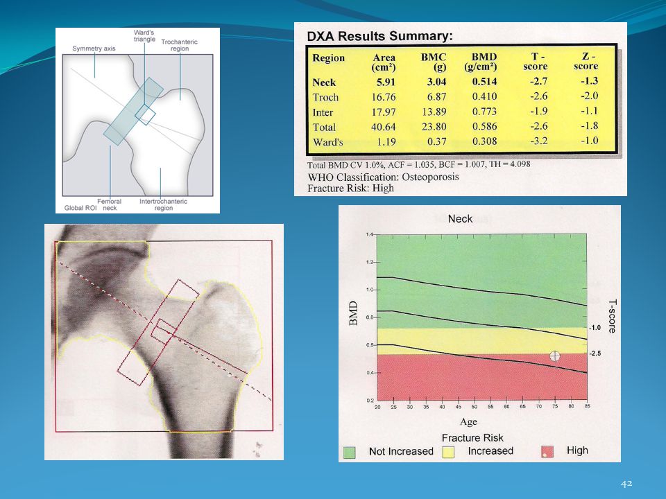
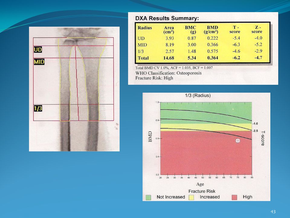
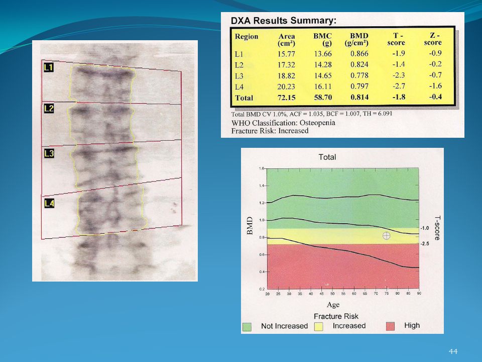
Definitions Bone mineral Density (BMD):the amount of mineralized tissue in the area scanned (g/cm²) T score: Compare your bone density to that of a young adult with normal bone density Z score: Compare your bone density to people of your age, gender, and ethnicity

45
Z-score Patient’s BMD – Age-Matched Mean BMD
1 SD of Age-Matched Mean BMD in g/cm2 เปรียบเทียบค่าที่วัดได้กับค่า predicted normal value ใน standard ที่อายุเดียวกัน

46
Calculation of z-scores
For example, a white woman aged 55 with BMD of 850 has a Z-score of ( )/139 = -0.18
/139 =")
47
T-score Example: 0.7 g/cm2 - 1.0 g/cm2 0.1 g/cm2 T-score = = - 3.0
Patient’s BMD – Young-Adult Mean BMD 1 SD of Young-Adult Mean BMD Example: T-score = 0.7 g/cm g/cm2 0.1 g/cm2 =

48
Diagnostic Classification
T-score Normal -1 or greater Osteopenia Between -1 and -2.5 Osteoporosis -2.5 or less Severe Osteoporosis -2.5 or less and fragility fracture

49
R2R R2R ย่อมาจาก Routine to Research หมายถึงการพัฒนางานประจำสู่งานวิจัย R2R จะทำให้การสร้างหรือการผลิตความรู้เกิดขึ้นได้ในทุกหนทุกแห่ง ทำให้ทุกที่เป็นองค์กรที่เรียนรู้ได้ด้วยตนเอง โดยใช้การวิจัยเป็นเครื่องมือแห่งการเรียนรู้

50
ตัวอย่างงานวิจัย

51
วัตถุประสงค์ This study was performed to assess the short-and long-term performance of a 6-year-old Lunar to measure lumbar spine,fermoral neck and forearm BMD continuously over this period J Wells and PJ Ryan; 2000

52
วิธีการทดลอง Short term precision
was assed by 15 measurement of the aluminium Lunar phatom (Madison), WI on the same day, with repositioning between scans was calculated by 10 measurements of the Hologic spine phantome (Waltham, MA) J Wells and PJ Ryan; 2000
, WI on the same day, with repositioning between scans. was calculated by 10 measurements of the Hologic spine phantome (Waltham, MA) J Wells and PJ Ryan;")
53
วิธีการทดลอง Long-term precision
was calculated in the same manner, from measurements over 18 months using the Hologic spine phantom, which were acquit three in a week J Wells and PJ Ryan; 2000

54
ผลการทดลอง The short-term coefficient of variation
was 0.242% using the Lunar aluminium phantom was 0.299% using the Hologic spine phantom J Wells and PJ Ryan; 2000

55
ผลการทดลอง Over the 18 months
Shift from base line 2.16% caused by PMT failure Manufacturer’s QC was not detected because software would detecte value than 2.5% PMT failure New PMT J Wells and PJ Ryan; 2000

56
ผลการทดลอง J Wells and PJ Ryan; 2000

57
ตัวอย่างงานวิจัย

58
วัตถุประสงค์ To estimate and compare the effective dose from DXA examinations in children and adults using a consistent methodology. G.M. Blake et al. / Bone 38 (2006) 935–942
 935–942.")
59
วิธีการทดลอง Dose were measuremented using thermoluminescent dosimeters (TLDs) Phantom Effective dose was calculated by taking the average dose to each organ and multiplying by its ICRP Publication 60 tissue-weighting factor

62
ขอบคุณครับ

งานนำเสนอที่คล้ายกัน


 98.08% 100.02% จังหวัด.>")
 ปี 57 เกิด 3 จับ 2 ราย (66.67.00% ) คดีเท่ากัน ผลการจับกุมบรรลุเป้า ( เป้า 75.47 %)>")


 ได้รับ จัดสรร ( ล้าน บาท ) เบิกจ่าย ร้อย ละ / งบ จัดสร ร สำนัก ชลประทานที่ 13 1,164,64 0,305.>")
